

8
The Mystery of Life
The grand unifying theme of Leonardo’s explorations of the macro-and microcosm was his persistent quest to understand the nature of life. Over the years, as he studied, drew, and painted the flows of water and air, the rocks and sediments of the Earth, the growth patterns of plants, and the anatomy of the human body, he correctly identified several of life’s key biological characteristics.
Early on, he recognized the fundamental role of water as life’s medium and vital fluid, the matrix of all organic forms (see p. 18). “It is the expansion and humor of all living bodies,” he wrote in one of his earliest Notebooks, Manuscript C. “Without it nothing retains its original form.”1 He associated the fluidity of water with the fluid and dynamic nature of living forms. He was especially fascinated by water vortices and other forms of turbulence, recognizing them intuitively, as I have argued, as symbols of life—stable and yet continually changing (see p. 22).
Nature as a whole was alive for Leonardo, and he saw similar patterns and processes in both the macrocosm of the living Earth and the microcosm of an individual organism. In view of this systemic approach—seeking to understand a natural phenomenon by linking it to other phenomena through a similarity of patterns—it is not surprising that Leonardo developed a conception of life that was deeply ecological. This is evident throughout his manuscripts, as, for example, when he describes the continual processes of growth and renewal that are common to all life on Earth (see p. 67):
Feathers grow on birds and change every year; hairs grow on animals and every year they change…. Grass grows in the fields and leaves on the trees, and every year they largely renew themselves.2
FACING The fetus in the womb, c. 1510–12 (detail, see plate 9).
Leonardo understood that these cycles of growth, decay, and renewal are linked to the cycles of life and death of individual organisms:
Our life is made by the death of others. In dead matter insensible life remains, which, reunited to the stomachs of living beings, resumes sensual and intellectual life…. Man and the animals are really the passage and conduit of food.3
With these statements, he anticipated the concepts of food chains and food cycles that would become the central focus of ecologists more than four hundred years later.4 It is also noteworthy that the phrase “life … sensual and intellectual” in this passage shows, like many other passages in the Notebooks, that Leonardo’s concept of life included its cognitive as well as its biological dimensions.
Leonardo recognized that the energy driving the ecological cycles of growth and renewal, of life and death, flows from the sun. In his studies of plant growth he noted: “The sun gives spirit and life to the plants, and the earth nourishes them with moisture” (see p. 119).5
Finally, Leonardo understood that both plants and animals need the surrounding air to sustain themselves. In Manuscript G he noted that the branches of trees “take in the air which nourishes them” (see p. 120);6 and in the Codex Atlanticus we find the observation:
Where the air is not in the right proportion to accommodate the flame, there no flame can live, nor any terrestrial or airborne animal…. Where the flame does not live, no breathing animal can live.7
The critical role of water as the matrix and nourishing fluid of living tissues, the life-sustaining role of air (or its oxygen, as we know today), the continual growth and renewal of all organic forms, the cycles of life and death in the natural world, and the life-giving power of the sun were the fundamental characteristics of life that Leonardo observed and analyzed. He explored them in the macrocosm in his studies of fluid dynamics, geology, and botany; and late in his life he began to examine the same patterns and processes in the microcosm of the human body, recognizing them as components of a system of metabolic processes that are understood in modern science as key characteristics of biological life.
In his explorations of the nature and origin of life in the human body, Leonardo focused on three interdependent processes. The first was the generation and transportation of the body’s “vital spirits” which, according to the ancient philosophers, arose from a mixture of blood and air (identified in modern biochemistry as oxygenated blood). The second process was the ebb and flow of breath in the lungs, and the third was the digestion of food and the transport of nutrients to the bodily tissues by the blood.
Leonardo recognized that these interdependent processes are essential for sustaining life. He also realized that at their very core was the human heart, and he hoped that detailed investigations of the nature and actions of this mysterious organ would bring him closer to understanding the mystery of life.
The Human Heart
Throughout the ages, the heart has been the bodily organ that has served as the foremost symbol of human existence and emotional life. We metaphorically associate the heart with a variety of emotions. We “hold someone in our heart” (love) and speak of “a kind-hearted person” (compassion); we “take something to heart” (seriousness); we thank someone “from the bottom of our heart” (sincerity); we don’t “have the heart” for a certain action (courage); and we make decisions “light-heartedly,” “with a heavy heart,” or after “a change of heart” (emotional depth).
In Leonardo’s time, the associations of the human heart with life, consciousness, and emotion were much more than just metaphors. From antiquity to the Middle Ages, the heart had been considered a unique organ that generated the body’s vital spirits (a mysterious “cardiac vapor”), as well as being the source of the body’s heat.8 For Aristotle (who was not aware of the central nervous system) the heart was not only the body’s center of vitality but the very seat of the soul, that is, of intelligence, motion, and sensation. Galen, the leading medical authority in antiquity, emphasized the “noble nature” of the heart and maintained that, even though it might look like a muscle, it was something entirely different. It circulated the vital spirits throughout the body together with its “innate heat”; its expansion and contraction, for Galen, were signs of its role as an intelligent organ. Avicenna, the great physician and philosopher, attempting to integrate Aristotle’s anatomy with Galen’s physiology, saw the heart as the body’s central and most important organ, but he stated that, being intelligent, it could delegate certain functions to other organs, especially to the brain.
Qui non estima la vita non la merita. (Ms. I, folio 15r)
One who does not respect life
does not deserve it.
Leonardo’s principal medical authority was Mondino, through whom he became acquainted with the works of Galen and Avicenna (see pp. 144–45). He accepted many of their concepts but readily departed from them when his observations taught him otherwise. Faced with this bewildering array of ideas about the heart inherited from antiquity, Leonardo concentrated on the twin problems, as he saw them, of how the actions of the heart maintained the blood at body temperature and how they produced the vital spirits that keep us alive. He adopted the ancient notion that these life-giving vapors arise from a mixture of blood and air—which is essentially correct, if we identify them with oxygenated blood—and he developed an ingenious theory to solve both problems.
As he did so often in his scientific investigations, Leonardo developed several theoretical models to explain the generation of body heat, discarding each model in turn when he found it unsatisfactory. His earliest attempt arose from the comparison of the flow of blood with the flow of water in “veins” inside the Earth and the flow of sap in plants. As I have discussed, Leonardo assumed that these three processes were all maintained by the same external power—the life-giving heat of the sun (see p. 18).
The sun, Leonardo thought, raises the “humors” (vital fluids) inside the three bodies: the water veins nourishing the Earth’s vegetation, the sap nourishing the plant tissues, and the blood nourishing the tissues of the human body. Having been elevated to heights where they cool and condense, the fluids fall down again, only to be raised anew in continual circulation. After a few years, Leonardo realized that his analogy between the blood vessels inside the human body and water veins inside the Earth was too narrow, and eventually he reached a full understanding of the water cycle (see p. 29). As far as the movement of blood was concerned, he proceeded to develop a second model in which the heart acts like a stove, housing a central fire.
The idea of a cardiac “hearth” that generates the heart’s innate heat had already been proposed by Aristotle. Leonardo linked this idea with a corresponding model of the water cycle in which water was supposed to be raised in special caverns inside the Earth in a process of distillation, fueled by the Earth’s internal heat (see p. 28).
In the Codex Arundel, Leonardo made a small sketch showing the heart as a kind of furnace with inlet and outlet valves opening into separate chimneys, which represented the lungs. On a sheet in the Windsor Collection, he outlined a two-chambered heart with passages connecting the two chambers to the lungs, a clear analogy to his model of the oven.9
However, Leonardo soon became dissatisfied with the view of the heart as merely an oven containing the fire of bodily heat. As he made more detailed dissections of various parts of the heart, he became aware of their functions in regulating the flow of blood. Thus he embarked on creating his third, and far more sophisticated, model of the heart, using his understanding of turbulent flows of water and air and the role of friction to explain the origin of both the blood-air mixture of the vital spirits and the body’s temperature. Although this model has serious flaws from the point of view of modern cardiology, it includes meticulous and accurate descriptions and drawings of many subtle features of the structure and actions of the heart and of the flow of blood—pioneering achievements in human anatomy.
To assess the significance of Leonardo’s cardiac anatomy, it is useful to briefly review the modern understanding of the anatomy and physiology of the heart and blood circulation. The human heart is a pear-shaped structure made of a special muscle tissue and enclosed in a membranous sac. A wall of muscle (the septum) divides the heart into two cavities, each consisting of an upper chamber (atrium) and a lower chamber (ventricle), as pictured in figure 8-1.
Two types of blood vessels connect to the heart’s four chambers: arteries, which carry blood away from the heart, and veins, which return blood to the heart. The main arterial vessel, the aorta, branches into smaller arteries, which in turn branch into still smaller vessels carrying blood to all parts of the body. Within the body tissues, the vessels are microscopic capillaries through which gas and nutrient exchanges occur.
After these exchanges, the blood converges from the capillaries into a network of minuscule veins, which in turn form larger veins that converge into the vena cava, the body’s principal vein. The inferior vena cava is supplied by veins from the legs, the liver, and the kidneys. The superior vena cava receives blood from the head and neck.
In order to prevent the flow of blood from backing up, the heart is equipped with a series of valves at various openings: the tricuspid valve between the right atrium and right ventricle, the mitral valve between the left atrium and left ventricle, and several semilunar valves in the aorta and the pulmonary artery. During the cardiac cycle, these valves open and close in a precise rhythm.
The system of blood circulation consists of two distinct parts. The systemic circulation serves the entire body except for the lungs, while the pulmonary circulation carries blood to and from the lungs. In the systemic circulation, oxygenated blood from the lungs enters the left atrium via two pairs of pulmonary veins (one pair from each lung). When it is filled, the atrium contracts, sending the blood into the left ventricle. A large percentage of blood also enters the ventricle passively, without atrial contraction.
The powerful left ventricle then contracts, forcing the blood under great pressure into the aortic arch and on into the aorta. Three major arteries originate from the aortic arch, supplying blood to the head, neck, and arms. Other major arteries from the aorta supply blood to the kidneys, the spleen and liver, and the thighs and legs. At the periphery of the body, oxygen and nutrients diffuse into the tissue cells, while carbon dioxide (CO2) and various metabolic waste products diffuse in the opposite direction, from the tissue cells into the capillaries, to be carried back to the heart by the veins. On this pathway of systemic circulation, part of the blood passes through the small intestine, where it absorbs nutrients from digested food, and proceeds to the liver for further digestion and regulation of various substances needed by the body. Another portion of blood goes through the kidneys, where the metabolic waste products are filtered out.
At the end of the systemic circulation, the blood, now low in oxygen and high in CO2, begins the pulmonary circulation by entering the right atrium of the heart, from where it is pressed into the right ventricle. The right ventricle then contracts, forcing the blood into the lungs through the pulmonary arteries. In the lungs, the oxygen-poor and CO2-rich blood flows through a vast network of capillaries surrounding the lungs’ tiny air sacs. Oxygen from the inhaled air diffuses across the capillary membranes into the blood, where it binds to hemoglobin molecules in the red blood cells, and CO2 diffuses in the opposite direction to be exhaled. The oxygenated blood returns to the heart via the pulmonary veins, entering the left atrium to complete the cycle.
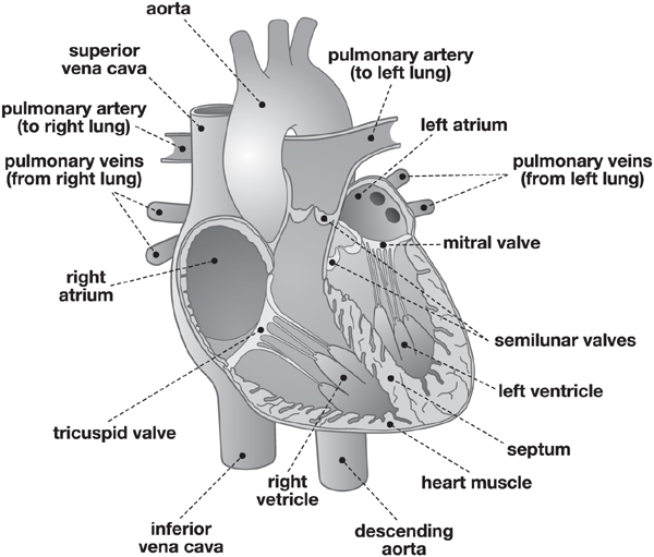
FIG. 8-1. The human heart and its blood vessels and valves.
In addition, there is a separate system of so-called coronary vessels to nourish the muscle tissues of the heart itself (not shown in fig. 8-1). Two coronary arteries originate at the base of the aorta and carry oxygen-rich blood to all the tissues of the heart through a delicate system of branching vessels. A corresponding system of coronary veins collects the oxygen-poor blood and delivers it to the right atrium.
The blood circulation is precisely synchronized with the cardiac cycle, or heartbeat, which consists of three phases. In the first phase, the two atria contract in symmetry, emptying their contents into the ventricles. A fraction of a second later, the two ventricles contract simultaneously, forcing blood into arteries that exit the heart. During the strong ventricular contractions, known as systole, the tricuspid and mitral valves snap shut, producing the familiar “lubb” sound, the first part of the heartbeat. Both atria and ventricles then relax briefly before the cycle repeats. At the beginning of the relaxation phase, known as diastole, the aortic and pulmonary valves (semilunar valves) close up, producing the characteristic “dubb” sound, the second part of the heartbeat. During the diastole, blood fills the atria and begins to flow passively into the ventricles. Both sides of the heart contract, empty, relax, and fill simultaneously. Therefore, only one systole and one diastole are felt.
It is noteworthy that in the pulmonary circulation, the arteries carry oxygen-poor blood (away from the heart) and the veins carry oxygen-rich blood (to the heart), whereas in the systemic circulation the oxygenated blood is carried by the arteries and the oxygen-poor blood by the veins. The pulmonary circulation intersects with the cycle of respiration in the lungs, while the systemic circulation intersects with the processes of digestion and waste excretion in the gastrointestinal tract, the liver, and the kidneys.
The Heart and Flow of Blood According to Galen
What Leonardo read about the structure of the heart and the flow of blood in the classical texts was quite different from our modern understanding.10 Galen conceived of the heart as primarily a respiratory organ, made of a special substance and endowed with unique properties. Through some mysterious processes, the heart produced both the innate heat of the body and the force of life, or “vital spirits,” and its most important function was to draw cool air from the lungs into the left ventricle, where the body heat was produced, to keep it from overheating.
This conception of the heart, which seems rather strange to us today, was derived from the fact that Galen’s entire physiology of the human body was based on the traditional idea of the soul as a fiery vital breath (pneuma), which took the form of several distinct “spirits” that were seen as the primary movers of all bodily functions.11
Galen postulated three of these life-giving forces. The “natural” spirits were created by the liver. They transformed digested food into blood and distributed it through the veins. The “vital” spirits were produced by the heart by transforming a portion of the air drawn from the lungs, and were then distributed through the arteries. And finally, the “animal” spirits, the body’s motor forces, originated in the brain and were transported to the muscles through hollow nerves.
Galen recognized only two chambers of the heart (the ventricles), viewing the atria merely as the endings of the vena cava and the pulmonary veins. He mentioned the cardiac valves but called them “orifices” and ignored their functions. Galen adopted the view of earlier Greek philosophers who had treated the arteries and veins as two completely separate systems, the former carrying the vital spirits (that is, essentially air) that were produced in the left ventricle of the heart, and the latter transporting the blood that was produced by the liver. However, Galen maintained that the arteries also contained some blood, which had passed through tiny pores in the septum from the right to the left ventricle.
Galen’s ideas about the movement of blood were rather confused. He believed that blood was made in the liver out of food, and that it carried nutrients to the bodily tissues through the veins (that is, in the opposite direction of the actual flow of blood in the veins). Some of that blood was sucked into the heart’s right ventricle when it dilated, and there it was “subtilized” (made thin and light). When the right ventricle contracted, some of this subtle blood seeped through the septum into the left ventricle, and the remaining blood passed to the lungs through the pulmonary artery to be exhaled together with air. In the left ventricle, the subtle blood combined with the air drawn from the lungs to form the vital spirits, which were then transported to the bodily tissues through the system of arteries.
However, in Galen’s theory the distribution of the vital spirits through the arteries, as well as that of the blood through the veins, was not by circulation but rather by fluctuations similar to the ebb and flow of the air in the lungs and the trachea. This must have been a natural assumption for him, since he thought of the heart as a respiratory organ and ignored the functions of the cardiac valves.
Leonardo’s Anatomy of the Heart
Leonardo’s sophisticated studies of the movements of the heart and blood, undertaken in Milan and Rome when he was in his early sixties, are the culmination of his anatomical work. He not only understood and pictured the heart like no one before him; he also observed subtleties in its actions and in the flow of blood that would elude medical researchers for centuries.
Leonardo illustrated his discoveries in a series of stunning drawings, now in the Windsor Collection. One of the most impressive is also one of his last anatomical drawings, dating from 1513. It is a magnificent double sheet showing the heart of an ox from several perspectives (plate 10). Leonardo’s main purpose in this study was to demonstrate the coronary vessels.12 In the two figures on the left side of the sheet, the coronary arteries are seen to originate at the base of the aorta and to divide into several branches. The pulmonary artery has been cut away so as to display the roots of the aorta and vena cava. The three cusps of the pulmonary valve are clearly visible in the orifice created by the severance of the artery.
The two figures on the top right show coronary veins as well as arteries. Their delicate branching patterns, crossing one another repeatedly, are beautifully rendered. The small sketches at the center of the right margin illustrate how the coronary vessels crown the heart, which explains their modern name. Below them, in the bottom right corner, Leonardo sketched the pulmonary valve and the tricuspid valve viewed from above, showing the latter both closed and open. The entire sheet is an impressive testimony to Leonardo’s understanding of many subtle features of cardiac anatomy.
The main part of Leonardo’s complex anatomical studies of the heart dates from the years 1511–13. At the beginning of this period, he recorded two major discoveries in a relatively large pocket book, now known as Manuscript G. The first was his conclusion that the heart, contrary to Galen’s view, is a muscle; the second was the observation that it had four cavities, not two, as all earlier medical authorities had believed.
On the very first folio of Manuscript G, next to several sketches of a dissected heart, Leonardo states categorically, “The heart is a principal muscle of force, and it is much more powerful than the other muscles.”13 After studying the classical medical texts in which the heart was said to be made of a special substance endowed with rather mysterious properties, and developing several theoretical models that failed to explain those properties, recognizing the heart as a muscle was a major breakthrough for Leonardo. Since he had already studied the nature and actions of muscles extensively, he realized that the heart, like any other muscle, had to be nourished by special blood vessels, and that its actions had to be triggered by special nerves. “The heart in itself is not the beginning of life,” he noted in the Anatomical Studies, “but is a vessel made of dense muscle, vivified and nourished by the artery and vein as are the other muscles.”14
During subsequent years, Leonardo explored the pathways and branching patterns of the coronary arteries and veins in great detail, summarizing the results of his investigations on the exquisite double sheet of the Windsor Collection (plate 10). At the same time, he looked for the nerves that stimulate the heart muscle and located them correctly in the large network known today as the vagi, or “wandering nerves.”* Leonardo called them “reversive nerves,” probably because of their frequent changes of direction, and in a note to himself emphasized the importance of their exploration:
Follow up the reversive nerves as far as the heart and see whether these nerves give movement to the heart, or whether the heart moves by itself. And if such movement comes from the reversive nerves, which have their origin in the brain, you will make it clear how the soul has its seat in the ventricles of the brain.15
It is evident from this passage that Leonardo correctly traced both the external and internal movements of the body back to the motor nerves and their origins in the brain.
Indeed, just as he represented the anatomy of the body’s muscles, tendons, and bones in terms of their movements, Leonardo pictured the heart in motion from the very beginning of his investigations. The sketches in Manuscript G are still quite imperfect, but already the heart is shown in action, with contracted or dilated chambers.
The fact that the heart has four cavities, rather than two, was the second major discovery of Leonardo’s early cardiac anatomy. He called the atria the “auricles of the heart”—a term still in use today—and he correctly identified their role as “the heart’s antechambers.”16 Leonardo was well aware of the importance of this discovery, repeating his assertion in several places in the Anatomical Studies.
Soon after his first sketches in Manuscript G, he produced a more elaborate drawing (fig. 8-2) in which the atria are clearly distinguished from the ventricles. In the accompanying text, Leonardo provides a succinct description:
The heart has four ventricles, that is two lower ones in the substance of the heart and two upper ones outside the substance of the heart. And of these, two are on the right and two on the left, and the ones on the right are much larger than the ones on the left. And the upper ventricles are separated from the lower ones by certain little doors, or gateways of the heart.17
On another folio, composed a couple of years later, he recorded a shorter version of the same statement: “The heart has four ventricles, that is two upper ones called auricles of the heart, and two below them called the right and left ventricle.”18
Leonardo fully understood the functioning of the cardiac valves, and he demonstrated with amazing accuracy their shapes in various stages of opening and closing.19 The characteristic H-shaped cleft of the mitral valve and the Y-shaped closures of the other three valves are clearly visible in several drawings of the cardiac orifices. The tricuspid valve, in particular, is explored in great detail, with respect to both its structure and its movements in action.20
Medical historian and Leonardo scholar Kenneth Keele has juxtaposed two of Leonardo’s drawings of the aortic valve in open and closed positions with two modern pictures of the same valve, obtained by Keele himself by means of high-speed photography (fig. 8-3). The result is stunning. The triangular shape of the orifice between the open cusps, the cusps’ wavy edges, and the detailed shape of the closed valve, as drawn by Leonardo five hundred years ago, are virtually identical to the anatomical features shown in the modern photographs.
In addition to demonstrating the precise shapes of the tricuspid and mitral valves in various positions, Leonardo dissected the so-called papillary muscles and showed how they contribute to controlling the actions of these valves by means of thread-like tendons, known today as chordae tendineae (“fibrous cords”). The way in which these fibers are attached to the valve cusps is demonstrated by Leonardo in great detail. It was not until the eighteenth and nineteenth centuries that the papillary muscles and their tendons were studied again with such meticulous care.21
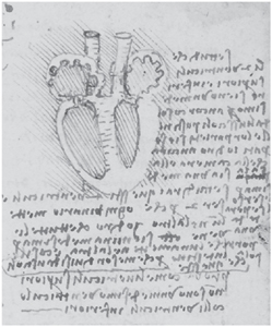
FIG. 8-2. The four chambers of the heart, c. 1511. Windsor Collection, Anatomical Studies, folio 155r (detail).
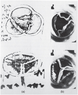
FIG. 8-3. Aortic valve cusps, open and closed, as seen in Leonardo’s drawings (a) and in modern high-speed photography (b). From Keele, Leonardo da Vinci’s Elements of the Science of Man, p. 319.
Leonardo’s precise representations of a variety of subtle anatomical structures of the heart were matched by his accurate descriptions of many cardiovascular functions. He correctly described the origin of the pulse as being in the rhythmic contractions of the arteries, which help the heart pump the blood and maintain a steady flow to the smaller vessels. He clearly recognized the connection between pulse and heartbeat:
The beating of the heart … generates a wave of blood through all the vessels, which continually dilate and contract…. And this we learn from the beating of the pulse when we touch the aforesaid vessels with the fingers in any part of the living body.22
Leonardo was also the first to appreciate that the heart shortens in systole (when it contracts) and lengthens in diastole (when it relaxes), which contradicted the traditional Galenic teachings and was confirmed 120 years later by the physician William Harvey. Even more remarkably, Leonardo offers the first correct interpretation of the cardiac impulse* against the chest wall:
The time of the contraction of the heart and of the percussion by its apex against the rib cage, of the beating of the pulse, and of the entrance of the blood into the front gateway of the heart [the aortic orifice] is one and the same.23
The realization that the contraction of the ventricles, the thump of the heart’s tip against the chest wall, the pulse, and the ejection of blood into the aorta all occur at the same time must be ranked as one of the greatest discoveries in Leonardo’s anatomy of the heart. None of his predecessors and contemporaries were aware that these phenomena are all interrelated. Again, it was Harvey who rediscovered their connections after more than a century had passed.
“Flux and Reflux” of the Blood
In view of Leonardo’s accurate visual demonstrations and verbal descriptions of so many subtle features of the cardiovascular system, it is difficult to believe that, unlike William Harvey, he did not recognize the circulation of the blood. Yet this is the case; so, why did Leonardo miss it and Harvey did not? Both of these brilliant scientists started from the same premise, the classical texts of Galen and Avicenna; both struggled with the same problems before the development of chemistry and the perfection of the microscope. Indeed, comparing Leonardo’s cardiac anatomy and physiology with those of Harvey is as revealing as comparing his mechanics with that of Galileo, Harvey’s contemporary (see p. 174).
One critical difference between the cardiac research of Leonardo and Harvey was that Leonardo refused to perform vivisections and therefore never saw the flow of blood through the heart and its vessels. He wrote about visiting an abattoir in Tuscany where he observed the slaughter of pigs and drew conclusions from the ways in which the blood gushed forth from their wounds, but performing vivisections himself was far too repugnant for Leonardo. Harvey, by contrast, would open up dogs and pigs, seemingly without any qualms, to observe and touch their “flagging” hearts when the animals died.24 In his accounts of these experiments, which sound quite cruel today, Harvey explained calmly and in gruesome detail how he obtained direct evidence about various aspects of the heart’s movements.
However, I believe that Leonardo might have recognized the circulation of blood even without evidence gained from vivisection. What prevented him from doing so much more fundamentally was the way in which he framed the whole issue of cardiac physiology. From his readings of the classical texts, Leonardo distilled two central questions: how does the heart generate the body heat that is characteristic of all mammals, and how does it maintain the life force, or “vital spirit,” that animates the bodily tissues? He used his sophisticated understanding of turbulent flows and of friction to develop a brilliant but erroneous model of blood flow, involving continual currents swirling back and forth between the heart’s atria and ventricles, thereby producing both the body’s innate heat and its vital spirits.
Leonardo’s outstanding discoveries in cardiac anatomy took him far beyond the prevalent views of Galen. He recognized the heart as a muscle exhibiting all the characteristics of muscular contraction; he identified four chambers of the heart instead of two; and he emphasized that the active movement of the heart was its contraction in systole, which expelled blood from the ventricles into the main vessels, rather than an expansion in diastole to draw air from the lungs into the heart, as Galen maintained. In fact, Leonardo disproved this Galenic view by inflating the lungs of a dead ox and observing that no air entered the pulmonary vein.25
In spite of all these advances, Leonardo clung to the fundamental Galenic idea that the blood moved in parallel in two separate vascular systems, and that this movement was one of ebb and flow—from the heart out to the body’s periphery and back to the heart—along both arteries and veins. There is no suggestion in Leonardo’s manuscripts of blood moving through tissue from the arteries to the veins, and hence no indication of any conception of circulation.
Leonardo maintained the Galenic conception of the ebb and flow of blood for several reasons. It was not contradicted by any of his observations, and it was consistent with his thorough studies of respiration—the ebb and flow of breath through the trachea and the lungs.26 He saw the ebb and flow of blood as another manifestation of the cyclical movements so characteristic of human physiology.
Most important, perhaps, was the fact that Leonardo postulated a cyclical movement of blood right inside the heart to explain the generation of body heat. As the atria and ventricles contract and dilate in synchrony, the blood flows back and forth between them, and by the friction in this “flux and reflux” it is heated and “subtilized.” This hydrodynamic model represented the very core of his cardiac anatomy, and it was fully consistent with the idea of cyclical movement of blood through the body. I think this was the main reason why Leonardo, with all his skillful anatomical dissections and great powers of observation, failed to recognize the blood’s circulation.
When he discovered the atria, he immediately had the idea of explaining the body’s heat as resulting from the friction of blood swirling through the four chambers of the heart. His extensive knowledge of turbulent flows allowed him to picture small vortices with great precision and to describe their motion accurately. On the very same folio of the Anatomical Studies that shows his first clear drawing of the atria and ventricles (see fig. 8-2), Leonardo provides a detailed description of his hydrodynamic model of heat generation:
The upper ventricles continually make a flux and reflux of the blood which is continually pulled or pressed by the lower ventricles from [or into] the upper…. And so, by such flux and reflux, made with great rapidity, the blood is heated and subtilized, and is made so hot that, but for the help of the bellows called lungs, which, by being dilated draw in fresh air, pressing it into contact with the coats of the ramifications of the vessels, refreshing them, the blood would become so hot that it would suffocate the heart and deprive it of life.27
Leonardo attempts here to provide a scientific explanation of body heat, which was seen by Galen as an innate and rather mysterious property of the heart. He knows that the principles of flow are the same for blood as for any other liquid (see p. 33), that friction always generates heat (see p. 202), and that liquids expand (become “subtilized”) when they are heated; and he assembles these observations from different areas of mechanics into a coherent (if incorrect) theoretical model.
Today we know that the body heat of mammals is the result of myriads of biochemical reactions throughout the bodily tissues, and that the body temperature is controlled by a heat-regulating center in the brain. Leonardo, living more than three hundred years before the development of biochemistry, could not have conceived of such an explanation. He erroneously assumed that the heat was generated by the swirling blood in the heart, but he was correct in his assumption that the blood is essential for distributing the heat throughout the body and maintaining a uniform body temperature. William Harvey, interestingly, did not attempt to explain the origin of body heat but simply assumed, without going into further details, that it was generated by the heart.28
Leonardo’s description of the role of the lungs in the passage quoted above is very intriguing. He adhered to the Galenic view that the function of the lungs was to cool the blood, but he contradicted Galen by asserting correctly that no air passes from the lungs to the heart. Therefore, Leonardo concluded, the cooling of the blood takes place when the air in the lungs “is pressed into contact with the coatings of the ramifications of the vessels.” This is a remarkably accurate description of the exchanges between the lungs’ air sacs and the network of blood vessels surrounding them—though what is exchanged, in Leonardo’s view, is not oxygen but merely heat.
On the subsequent folio, Leonardo continues his discussion of body heat with a detailed description of the turbulent flows of blood in the heart:
And so, between revolving up and down successively it never ceases to flow through the cavernous recesses interposed between the muscles which contract the upper ventricle. And the whirling round of the blood in diverse eddies, and the friction it makes on the walls, and the percussions in the hollows, are the cause of the heating of the blood, and making it from thick and viscous to subtle and penetrative, suitable for flowing from the right to the left ventricle through the narrow porosities of the wall interposed between that right and left lower ventricle.29
In addition to generating the body temperature, the heating of the blood serves two further purposes, in Leonardo’s view. On the right side of the heart, as he explains in the above passage, it is transformed from a “thick and viscous” liquid into one that is so “subtle and penetrating” that it can pass to the left ventricle through the invisible pores in the septum postulated by Galen. Harvey, in contrast to Leonardo, rejected the Galenic conception of invisible septal pores, postulating correctly that the blood moves from the right to the left side via the pulmonary circulation. However, Harvey was never quite comfortable with this argument, since it replaced the idea of blood seeping through invisible pores in the septum with that of its passage through equally invisible capillaries in the lungs.30
The Vital Spirits
Galen’s physiology of the human body, as I have mentioned, was based on three types of life-giving forces, or “spirits,” which were seen as different manifestations of the same vital breath (pneuma) and as the primary agents of all bodily functions (see p. 288). Leonardo, with his strictly empirical approach to scientific knowledge, which rejected all notions of supernatural forces,31 eliminated two of Galen’s spirits from his conception of human physiology. He questioned the ancient idea of animal spirits moving through hollow nerves as some kind of “psychic wind,” and replaced it with the much more sophisticated concept of immaterial nervous impulses traveling through the sensory and motor nerves in the form of waves.32 He also rejected the Galenic notion of natural spirits, the agents of digestion, as nonexisting entities for which he could find no evidence (see p. 309).
However, Leonardo retained Galen’s concept of vital spirits as a fundamental force of life. Having identified the life-sustaining role of air in numerous observations of animal and plant life (see p. 282), he conceived of the idea that these spirits were some vital essence of air, which was isolated in the heart, intermingled with blood, and then transported to the periphery of the body in order to animate all bodily tissues. From our modern perspective we can see that Leonardo’s intuition was absolutely right. Oxygen is the life-sustaining essence of the air, and oxygenated blood is the “mixture” of blood and air that animates the body’s tissues.
Leonardo’s theoretical model for the generation of the vital spirits is essentially the same hydrodynamic model he used to explain the generation of body heat, but the process is somewhat more complex and takes place predominantly in the left ventricle:
[The blood] is more heated in the left ventricle where the walls are thick than in the right ventricle with the thin wall. And that heat subtilizes the blood and vaporizes it, and converts some of it into air…. The lung cannot send air into the heart, nor is it necessary, because as said, air is generated in the heart [and] mixed with heat and condensed moisture.33
According to Leonardo, the blood-air mixture is not obtained from air sucked in from the lungs, as Galen believed, but is produced by vaporizing part of the blood. In this way, the vital spirits are formed out of heat, humidity, and the mixture of blood and air. To generate the necessary heat for this process, the turbulence of the blood in the left ventricle must be much stronger than that in the right. For Leonardo, this is confirmed by the fact that the muscle walls of the left ventricle are thicker than those of the right ventricle. (We know now that this is due to the greater force needed to pump the blood through the systemic circulation.)
Like his explanation of the body temperature, Leonardo’s account of the generation of the vital spirits integrates observations from several areas of his science into a coherent theoretical model: the relation between friction and heat, between air and the sustenance of life, and between heat and the body’s living tissues—or between energy and metabolic processes, as we would say in modern scientific language. As Leonardo sums it up, “[without] flux and reflux the blood would not be heated, and consequently the vital spirits could not be generated, and therefore life would be destroyed.”34
According to Leonardo, the vital spirits created in the left ventricle are “augmented and vivified” by further turbulences as the blood enters the base of the ascending aorta:
The revolution of the blood in the anteroom of the heart at the base of the aorta serves two effects, of which the first is that this revolution, multiplied in many aspects, makes within itself great friction, which heats and subtilizes the blood, and augments and vivifies the vital spirits, which always maintain themselves in warmth and humidity. The second effect of this revolution of the blood is to close again the opened gates [valve cusps] of the heart with its first reflected motion, with perfect closure.35
This passage summarizes what must be seen as the most sophisticated and most extraordinary piece of Leonardo’s anatomy of the heart. To determine the precise shape of the turbulences in the aorta, he analyzed the flow patterns behind the aortic valve in incredible detail, picturing them repeatedly from various angles (for example, fig. 8-4) and describing them in long paragraphs on several folios of the Anatomical Studies.36 He showed that the blood, as it passes through the valve’s triangular orifice, forms three distinct eddies in a retrograde motion after impinging on the stationary blood already in the aorta. Leonardo demonstrated that these vortices are generated when the blood flows through three pouches in the wall of the aorta right behind the aortic valve. He called these hemispherical pouches “hemicycles.” Today they are called the sinuses of Valsalva in honor of the anatomist Antonio Valsalva, who rediscovered them in the eighteenth century.
The most remarkable part of Leonardo’s analysis of the three vortices is his discovery that they fill out the semilunar cusps of the aortic valve and begin to close it before the ventricle’s contraction ends and the weight of the column of blood in the aorta shuts the valve completely. Leonardo finds a persuasive argument against the valve being closed by the weight of the blood above it alone: “The shape of the valve denies this, as it would quicker be crushed than shut,” he writes next to a tiny sketch of a crushed valve cusp (see fig. 8-4, top left corner).37

FIG. 8-4. Turbulent flows of blood at the base of the aorta, 1513. Windsor Collection, Anatomical Studies, folio 172v (detail).
Leonardo realized that determining the exact patterns of turbulence in the aorta was extremely difficult, and sometimes he was doubtful about whether his interpretation was correct. “Such doubts are subtle and difficult to prove and clarify,” he mused.38 But he did not leave things there. Incredible as it may seem, especially in view of his advanced age at the time, Leonardo planned to test his hypothesis by building a glass model of the aorta’s base, including the aortic sinuses, with a valve taken from the heart of an ox. He would pour water into the model, with millet grains mixed in, to observe the turbulent flows.* “But first pour wax into the gate of the heart of an ox,” he reminded himself, referring to a technique of dissection he had used many years earlier, “so that you may see the true shape of this gate.”39
We do not know whether Leonardo ever built his glass model of the aorta. In any case, the experimental verification of his hypothesis (that the closure of the aortic valve is initiated by eddies of blood swirling through the aortic sinuses) had to wait for more than four hundred years.40 This is certainly one of the most astonishing cases of a scientific discovery far ahead of its time. Here is how medical historian Sherwin Nuland tells the story.
Until at least the early part of the twentieth century, it was assumed by all cardiac researchers that the valve between the heart and the aorta (the aortic valve) functions passively, like that of a standard water pump: When the heart contracts, it pushes blood out and forces the valve open so that the blood can be ejected upward into the aorta; when the pressure of contraction lessens, the valve is forced shut by the weight of the column of blood in the aorta, pressing down from above it. This seemed a perfectly straightforward explanation of the hydraulics of the system….
But in 1912 it was demonstrated that the dynamics are not quite as simple as had been thought. In fact, the process of valve closure was shown to be somewhat more gradual than could be accounted for by an abrupt change in pressure…. Decades more had to pass before investigative technology had reached such an advanced state that the details could be satisfactorily explored and actually visualized. By the 1960s, dye and cineradiography methods had been sufficiently developed that it was possible to study flow patterns with extreme accuracy. It was demonstrated that some of the blood which is ejected into the aorta swirls into the [sinuses of Valsalva] and forms eddy currents that exert pressure on the upper surface of the valve, causing it to begin closing even before the ventricle has completed its contraction. This could not have been known without the new research methods.
Or so it was thought. Leonardo da Vinci had shown the same thing in the first decade of the sixteenth century…. Both his text and illustrations clearly show the correct mechanism of both opening and closure of the three leaflets that make up the aortic valve, including the fact that the initiation of the closure is due to eddy currents originating in the sinuses of Valsalva. He demonstrated repeatedly that the closure is gradual. Leonardo’s observations are identical with those that would be made by groups of researchers in a series of studies beginning in 1969, and he drew the same conclusions from them as they would.… Of all the amazements that Leonardo left for the ages, this one would seem to be the most extraordinary.41
The Flow of Blood Through the Body
When Leonardo visualized the flow of blood through the chambers of the heart and through the arteries and veins, his main concern was to understand how the vital spirits are generated, how they “vivify” the bodily tissues, and how the blood nourishes these tissues. In other words, he wanted to understand the body’s basic metabolic processes (as we would say today)—the very essence of biological life.
The precise mapping of the movement of blood through the heart and body was a secondary issue for Leonardo. As medical historians O’Malley and Saunders point out, his views on the subject varied considerably over time, and he never recorded them in a complete statement.42 According to these scholars, Leonardo’s theory of the movement of blood may be summarized as follows.
At the beginning of the cardiac cycle, the right atrium contracts, while the right ventricle dilates, which makes the blood rush into the ventricle, thus creating turbulent currents. Then the right ventricle contracts, while the atrium dilates, sending blood back to the atrium and also into the lungs through the pulmonary arteries. A small portion of “subtilized” blood seeps through the septal pores into the left ventricle. This process is facilitated by the dilation of the left ventricle during the contraction of the right. During its dilation, the left ventricle also receives blood from the contracting left atrium.
Then the left ventricle contracts, sending part of the blood back to the atrium and ejecting the other part through the aorta. In the forceful flux and reflux between the left atrium and ventricle, the blood is heated considerably and some of it is vaporized to generate the vital spirits, which are sent to the bodily tissues through the aorta together with the ejected blood. A small part of the vaporized blood enters the lungs through the pulmonary veins and escapes into the bronchi. On both sides, the blood is cooled in the lungs before returning to the atria and ventricles.
In Leonardo’s cardiac cycle, the atria and ventricles contract and dilate alternatively, as in the modern understanding of blood circulation. However, the ventricles on the left and right do not contract in symmetry, so as to facilitate the passage of blood through the septal pores when the right ventricle contracts while the left expands. In other words, the right atrium and left ventricle contract in symmetry during the first phase of Leonardo’s cardiac cycle; and the left atrium and right ventricle contract in symmetry during the second phase.
One problem of this scheme was to explain how the blood could pass back and forth between the atria and ventricles through the valves that separate the two chambers on both sides of the heart. Leonardo tackled this problem by proposing a complex sequence of synchronized actions of the two valves through rhythmic contractions and relaxations of their papillary muscles. As Keele commented, “It is a very neatly reasoned and consistent account of papillary muscle action. Its error lies basically in Leonardo’s ignorance of the fact that the papillary muscles are part of the main mass of cardiac muscles, all of which contract in systole.”43
As far as the flow of blood through the arteries and veins was concerned, Leonardo retained the fundamental Galenic idea of its ebb and flow in two separate vascular systems, as I have mentioned. This implied that once the blood reaches the body’s periphery, it is used up by the tissues for their “vivification” and nourishment, which means that it has to be constantly replenished. Following Galen, Leonardo assumed that the blood was formed in the liver, which is not completely wrong, since some essential components of blood (including blood-clotting substances and other plasma proteins) are indeed manufactured there.
In his dissections of blood vessels, Leonardo’s starting point was the works of Avicenna and Mondino (see pp. 144–45). In both of these texts, there is considerable confusion between arteries and veins, some of which is still present in Leonardo’s work when he uses the term vene indiscriminately to describe either arteries or veins. Leonardo produced most of his drawings of blood vessels several years before his sophisticated anatomies of the heart. They reached their climax with his dissection of the centenarian around 1508, which he documented with a series of magnificent anatomical drawings (see p. 227).
One of the most accomplished of these anatomical studies shows the blood vessels of the liver, known today as the portal and hepatic vessels (fig. 8-5). The major branches of these arteries and veins, as well as the fractal structures of their ramifications in the body of the liver, are depicted by Leonardo with such clarity that modern medical scientists can easily identify them.44
Blood is carried to the liver via two large vessels. The hepatic artery carries oxygen-rich blood from the abdominal aorta, and the portal vein carries blood containing digested food from the gastrointestinal tract. These blood vessels subdivide repeatedly in the liver until they form minute capillaries through which the blood enters into clusters of hepatic cells, known as lobules. The liver tissue is composed of thousands of these lobules, in which the nutrients transported by the blood are further digested and various toxic substances are filtered out. Having thus been cleansed, the blood is collected by a corresponding network of minuscule veins that converge into progressively larger veins, eventually ending up in the single hepatic vein that drains the blood into the vena cava.
The drawings in figure 8-5 show that Leonardo has dissected the blood vessels out of the liver substance in order to clearly demonstrate their branching patterns. The figure on top of the page shows the networks of blood vessels formed in the liver by the hepatic artery and the portal vein. Leonardo shows how the artery arises from the abdominal aorta and how it gives off several branches that carry blood to the other digestive organs before it enters the liver.*
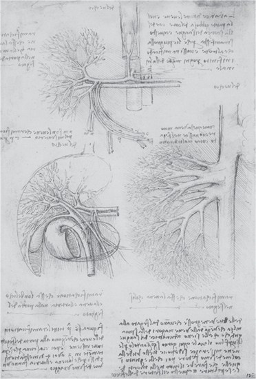
FIG. 8-5. The blood vessels of the liver, c. 1507–8. Windsor Collection, Anatomical Studies, folio 60v.
Below the branches of the hepatic artery lie the portal vein and its ramifications (the hepatic veins). Both the hepatic artery and the portal vein are seen to divide into right and left branches in the liver before generating a sequence of smaller and smaller ramifications. The large network of hepatic veins with its numerous branches, all draining into the vena cava, is demonstrated in the lower right figure. In addition, Leonardo loosely sketched the opening of the vena cava into the right atrium of the heart.
The figure on the left is partly the same as that above. But here Leonardo has indicated the outlines of the liver and stomach, and he has added some of the vessels, or “ducts,” that carry bile from the liver to other digestive organs. In the words of O’Malley and Saunders, “The gall bladder, cystic duct, hepatic ducts and common bile duct passing to the duodenum are amazingly portrayed.”45 The fact that medical scientists today have no problem identifying these anatomical details, which are bewildering for the lay person, is impressive testimony to Leonardo’s anatomical skills, holistic memory, and mastery of pictorial demonstration.
Leonardo performed his anatomy of the centenarian, who died in his presence without having experienced any major illness, “in order to see the cause of so sweet a death” (see p. 227). His careful dissection of the old man’s entire body resulted not only in a series of superb studies of the blood vessels and internal organs, but also in Leonardo’s most famous and most amazing medical discovery.
He observed that the blood vessels of the old man had thickened with age and that their ramifications had become tortuous. This observation was reinforced when he had the opportunity, by coincidence, to dissect the smooth and straight vessels of a two-year-old child around the same time. Leonardo recorded his observations on a folio of the Anatomical Studies with two small sketches, one showing a straight set of branching vessels labeled “young,” and the other a set of twisted ramifications labeled “old” (fig. 8-6). In a note next to the drawings, under the heading “Nature of the vessels in youth and old age,” Leonardo succinctly describes the condition now known as arteriosclerosis:
FIG. 8-6. Blood vessels of the young (right) and the old (left), c. 1507–8. Windsor Collection, Anatomical Studies, folio 69r (detail).
In so far as the vessels become old, their straightness is destroyed in their ramifications, and they become so much more sinuous, or twisting, and of a thicker coat, as their age is fuller in years.46
On the verso of the same folio, Leonardo develops the theme of arteriosclerosis with age at some length, together with his brilliant and correct interpretation of the condition, which he observed to be particularly severe in the blood vessels supplying the centenarian’s liver. Leonardo begins his analysis with a description of the thickening of the vascular walls, due to their prolonged contact with the nourishing blood, the consequent obstruction of the blood flow, and the effects of the diminished blood supply on the liver:
The artery and the vein … acquire so thick a skin that it restricts the passage of the blood…. And these vessels, apart from the thickening of their skin, grow in length and twist like a snake, and the liver loses the nourishment of the blood which was carried there by that vein. Thus the liver becomes desiccated and like congealed bran both in color and in substance, so that when it is subjected to the slightest friction, its substance falls away in small particles like sawdust and leaves behind the veins and arteries.47
According to O’Malley and Saunders, this passage contains not only the first description of arteriosclerosis in medical history, but also the first vivid and clear account of cirrhosis of the liver.48 A couple of paragraphs below, on the same page, we find Leonardo’s moving story of his encounter with the old man at the hospital of Santa Maria Nuova, followed by his diagnosis of the centenarian’s cause of death:
And I made an anatomy of him in order to see the cause of so sweet a death, which I found to be weakness from lack of blood and deficiency of the artery [aorta] that nourishes the heart and other parts below it, which I found very dry, thin, and withered…. The other anatomy was that of a child of two years in which I found everything to be the opposite to that of the old man.
In the margin of the same page, Leonardo concludes his analysis by summarizing arteriosclerosis in general terms:
The aged who enjoy good health die through lack of nourishment. This happens because … the passage of the meseraic [portal] veins is continually constricted by the thickening of the skin of these veins, progressing as far as the capillary vessels which are the first to close up entirely.
This passage is noteworthy also because it shows that Leonardo was the first to use the term “capillary vessels” (vene capillari) for the minute blood vessels, which were unknown in his time.
Finally, at the end of the page, Leonardo puts his discovery into the overall context of his science of living forms by observing that the aging process of blood vessels represents a general pattern of behavior of living tissues:
[The] coat of the vessels acts in man as in oranges, in which, as the skin thickens, so the pulp diminishes the older they become.
After Leonardo’s detailed description and correct interpretation of arteriosclerosis, it took another three hundred years for the condition to be rediscovered. In the early nineteenth century, the anatomist Antonio Scarpa accurately described and illustrated the thickening of the arterial walls, based on his dissections, but attributed the process merely to “internal unknown causes.” Some thirty years later, the pathologist and surgeon Jean Lobstein coined the term “arteriosclerosis” and, like Leonardo, asserted that the condition was due to an “abnormal state of nutrition” of the tissues.49 The role of cholesterol and other fatty substances in the thickening and hardening of the blood vessels was investigated only during the second half of the twentieth century and is still not fully understood today.
The Digestive System and the Body’s Metabolic Processes
When Leonardo investigated the blood supply to the liver and reflected on the effects of the nutrients in the blood on the arterial walls, he had already carried out extensive studies of the entire digestive system.50 During the three years preceding his dissection of the centenarian, 1506–8, he produced a series of splendid representations of the abdominal muscles, as well as the stomach, intestines, liver, and spleen. Moreover, he demonstrated the blood supply to the gall bladder and depicted the intricate intestinal blood vessels. Leonardo was the first to show the correct relative positions of the small and large intestines, and the first to recognize and clearly depict the appendix.
In his investigations of the physiology of digestion, Leonardo accurately described the act of swallowing and the passage of food through the esophagus to the stomach and the intestines. However, he was unaware of peristalsis, the waves of contractions of the intestine that pass along the food, having never observed it because of his ethical objection to vivisection. Leonardo attributed the movement of food through the digestive tract to pressure exerted by the diaphragm in its downward movement. A folio of the Anatomical Studies contains a detailed discussion of the diaphragm’s dual function* as “the motor of food and air.”51
Without access to biochemistry, Leonardo explained the process of digestion that we now associate with digestive enzymes by assuming that the food was broken down and liquefied in the stomach and intestine by bodily heat in a process of “coction.” This was consistent with the Galenic teachings, but Leonardo disagreed with Galen’s view that the heat required for digestion arose spontaneously in the intestine. Instead, he postulated that it was conveyed to the digestive tract by the blood in the intestinal arteries.
Leonardo correctly observed that the partially digested nutrients are carried to the liver in the portal vein. He assumed with Galen that in the liver they were transformed into blood, and he also recognized that various impurities were eliminated from the blood to be excreted with the urine. However, he erroneously saw the bile (which facilitates the digestion and absorption of fat) as a collection of waste products, stored in the gall bladder to be excreted.52 Leonardo’s errors are hardly surprising; any explanation without the help of chemistry was bound to be woefully inadequate.
Having analyzed the entire process of digestion from the mastication and swallowing of food to its transformation into blood and the excretion of waste products, Leonardo turned his attention to the metabolic processes at the body’s periphery. Without a microscope, he could not observe the exchanges of nutrients, wastes, and “vital spirits” (oxygen) between the tissues and capillaries, but he showed remarkable intuition in postulating them. In fact, he intuited a fundamental aspect of tissue metabolism (to use the modern scientific term) that would be rediscovered only in the twentieth century.
Leonardo recognized three important functions of the blood: to “vivify” the tissues by supplying them with oxygen (the “vital spirits” generated in the heart), to nourish them with the nutrients absorbed from digested food, and to carry away broken-down tissues and wastes.53 The vital spirits, according to Leonardo, dissolve in the capillaries; their life-giving essence (identified today with oxygen) is absorbed by the tissues, and the remaining hot moisture “evaporates through the terminations of the capillary vessels at the surface of the skin in the form of sweat.”54 Leonardo’s chemistry is of course rather fuzzy here, and he did not realize that the blood, having delivered the oxygen to the tissues, continues its circulation by draining into a network of veins. Nevertheless, his intuitive understanding of the diffusion of oxygen from the capillaries to the tissues is remarkable.
Similarly, Leonardo correctly intuited the exchange of nutrients and waste products between the capillaries and tissues, and it was this intuitive understanding that led him to one of his most extraordinary discoveries. He had previously compared the life-sustaining role of air for animals to the way air sustains the “life” of a flame (see p. 282). Now he extended this analogy to the nourishment of the bodily tissues by the blood in a long passage, titled “how the body of an animal continually dies and is reborn”:
The body of anything that is nourished continually dies and is continually reborn, for nourishment cannot enter except into those places where past nourishment has been exhausted; and if it has been exhausted it no longer has life. If you do not supply nourishment equal to the nourishment that has departed, then life fails in its vigor; and if you take away this nourishment, life is totally destroyed. But if you supply just as much as is destroyed daily, then as much of the life is renewed as is consumed, just as the light of the candle is made from the nourishment it receives from the liquid of that candle.55
The brilliant intuitive insight expressed by Leonardo in this passage is that every living organism, like a burning flame, needs to feed on a continuous flow of nutrients to stay alive, and that these nutrients are continually absorbed and transformed by the body’s living tissues while waste products are generated in the same process. Leonardo continues the passage by elaborating his analogy between the continual stream of death and renewal in living tissues and the process of combustion in the flame of the candle:
And this light is also continually renewed with the swiftest assistance from below by as much as is consumed above in dying, and in dying the brilliant light is converted into murky smoke. This death is continuous as long as the smoke continues, and the continuity of the smoke is equal to the continuing nourishment; and in the same instant the whole light dies and is completely regenerated together with the motion of its nourishment.
With this insight, described precisely and in beautiful poetic language, Leonardo was centuries ahead of his time. The definition of living organisms as “open systems” that need to feed on a continual flux of energy and matter was proposed by the biologist Ludwig von Bertalanffy in the 1940s, more than four hundred years after Leonardo had clearly recognized it. “The organism is not a static system closed to the outside and always containing the identical components,” Bertalanffy wrote. “It is an open system in a (quasi-) steady state … in which material continually enters from, and leaves into, the outside environment…. The fundamental phenomena of life can be considered as consequences of this fact.”56
It took another thirty years before a mathematical theory of open systems was formulated. This was achieved by physicist and chemist Ilya Prigogine, who called those systems “dissipative structures” and was awarded the Nobel Prize for his theory.57 Today, the flow of energy and matter through a dissipative structure is considered a defining characteristic of life, and the image of the flame of a candle is still used as a simple illustration.
The Nature of Life
Leonardo’s sophisticated studies of the heart and the flow of blood, undertaken in old age, represented the climax of his anatomical research and, at the same time, led him to some of his deepest insights in his long-standing quest to understand the nature of life. He recorded meticulous descriptions of many subtle features of cardiac physiology—including the coordinated actions of the heart’s four chambers (when his contemporaries knew of only two), and the corresponding synchronized actions of its valves—and he illustrated them in a series of superb drawings. According to Keele,
Leonardo’s success in cardiac anatomy [is] so great that there are aspects of the work which are not yet equaled by modern anatomical illustration…. His consistent practice of illustration of the heart and its valves, both in systole and in diastole, with a comparison of the position of the parts, has rarely if ever been performed in any anatomical textbook…. His detailed treatment of the valves and their movement is such that it is difficult to find illustrations in modern books with which to compare them.58
Leonardo missed some crucial details of the mechanics of blood circulation because of his pursuit of the forbidding challenge to explain (without chemistry) the generation of the body’s temperature and of the blood’s life-giving essence, identified today with oxygen. At the same time, this focus on metabolic processes enabled him to recognize many fundamental features of tissue metabolism that would be rediscovered only centuries later. These include the insights that heat energy supports the metabolic processes; that oxygen (the “vital spirits”) sustains them; that there is a constant flow of oxygen from the heart to the tissues at the body’s periphery; that the blood returns from these tissues carrying metabolic waste products; and, finally, what must be seen as Leonardo’s crowning achievement, the insight that the continuous absorption and transformation of nutrients, with concomitant generation of waste products, is a necessary condition for sustaining life and hence a fundamental property of all living tissues. It is evident from these achievements that Leonardo’s theory of the functioning of the heart and the flow of blood led him to understand some of the defining characteristics of biological life.
The Origin and Early Development of Human Life
While Leonardo immersed himself in studying the subtleties of the movements of the heart and the flow of blood, he became intensely interested in another dimension of the mystery of human life—its origin and unfolding in the processes of reproduction and embryonic development. The main body of Leonardo’s embryological studies is represented on three Windsor folios (196, 197, and 198) dating from 1510 to 1512. These folios were initiated during his last years in Milan, but in Rome he added several important notes and smaller sketches.59
In Leonardo’s time, there was a lively debate among philosophers as to whether the human embryo was formed from two “seeds” (one from the male parent and the other from the female), or whether its characteristics derived from the male seed only. Aristotle taught that all inherited characteristics came from the father, with the mother providing merely the nutritive bed in which the embryo could grow. Galen, by contrast, believed that both the father’s and the mother’s “seeds” contributed to the embryo’s characteristics. After examining both theories, Leonardo unequivocally sided with the Galenic view, offering the following empirical support for it.
If a black man impregnates a white woman, she will give birth to a grey child, and this shows that the seed of the mother has equal power in the embryo to the seed of the father.60
The way Leonardo exposed Aristotle’s ideological bias with this simple observation is typical of his assured empirical approach to knowledge.
FIG. 8-7. Early embryo (1–2 months). Codex Atlanticus, folio 1064r (detail).
Although Leonardo performed at least one anatomy of a human fetus,61 most of his embryological studies were based on dissections of cows and sheep. The earliest stage of embryonic development he recorded is in a small sketch in the Codex Atlanticus. It shows an oval embryonic body surrounded by amniotic fluid and attached to the uterine wall by a bodystalk (fig. 8-7). According to Keele, an embryo of that appearance, whether animal or human, can be dated as being one to two months old.62
Leonardo observed the same structure in the ovaries of flowering plants, where the seed, during its first stage of development, remains attached to the ovarian wall by a stalk. As I have discussed, he viewed this stalk as the equivalent to the umbilical cord, as botanists do today.* This observation, recorded on one of the folios of embryological studies in the Windsor Collection,63 is an impressive testimony to Leonardo’s ability of seeing similar patterns in different living systems.
FIG. 8-8. Study of an early embryo. Codex Atlanticus, folio 313r (detail).
Another rough sketch from around the same time shows a fetus in utero in the second month surrounded by a series of membranes (fig. 8-8). In an accompanying sketch below, Leonardo shows the appearance of the fetus after he has extracted it and has sliced the membranes, opening them like flower petals.
The most sophisticated and famous of Leonardo’s embryological drawings depicts a five-month-old fetus as seen through the incised uterine wall (plate 9). The positions of its head and limbs are rendered with an accuracy never seen before and, one feels, with awe and reverence for the unfolding of human life.
Below and to the right of the main drawing, Leonardo shows the stages of his dissection, as he delicately slices and peels off the fetal membranes. Again, these images are strikingly reminiscent of the petals of a flower or the outer layers of a seed—moving visual expressions of Leonardo’s deep conviction of the unity of life at all scales of nature.
At the center of the right margin we find an intriguing diagram of a ball rolling up an inclined plane. In the accompanying note, Leonardo explains that it can do so because a piece of lead weighing more than the rest of the ball is attached to its periphery at the uphill end. This diagram provides a concise mechanical explanation of how the weight of the head of the fetus enables its body to rotate within the uterine “ball of water” right before birth, so as to engage head first.
Although Leonardo’s representation of the fetus in the womb is obviously based on the dissection of a pregnant woman after her unfortunate death, the uterine membranes and placenta, as shown in figure 8-9, are those of a cow. To experts in anatomy this is evident from the stylized rendering of the placenta as a series of individual patches attached to the fetal membrane. These placental patches, known as cotyledons, are typical of the uterus of cows.
In the sketches at the top of the page of plate 9, Leonardo studies in great detail how the protuberances of the fetal membranes, known today as villi, interlace with corresponding projections from the uterine wall. Here too, the interdigitation of the placenta and uterine wall is shown to occur in the distinct patches (cotyledons) typical of cows.

FIG. 8-9. The fetal calf in utero, c. 1506–8. Windsor Collection, Anatomical Studies, folio 52r.
Leonardo dissected animals not only to find anatomical analogies to the human body but also to explore the animal body in its own right. Among his anatomical studies in the Windsor Collection there is a sheet with two superb illustrations of the pregnant uterus of a cow (see fig. 8-9). The upper drawing shows the crescent-shaped womb with the uterine and ovarian blood vessels. In the drawing below, as Keele explains, Leonardo has removed the uterine wall to expose the scattered cotyledons and reveal the fetus through the inner transparent membranes.64 The fetal calf, lying in the womb upside down with its head to the left, its forelegs and cloven hoofs on top, and its body stretching across to the hind legs on the right, is rendered with the same delicacy and care as the human fetus. In fact, Leonardo’s “transparency technique” is so subtle that the calf’s body is often not recognized at first viewing.
Leonardo’s embryological drawings are graceful and touching revelations of the mysteries surrounding the origins of life. They epitomize the artist’s great care, sensitivity, and tremendous respect for life, both animal and human, and exude a tenderness that is deeply moving. In the words of physician Sherwin Nuland,
[His] depiction of a five-month fetus in the womb is a thing of beauty…. It stands as a masterwork of art, and, considering the very little that was at the time understood of embryology, a master-work of scientific perception as well.65
In addition to studying the anatomies of human and animal embryos in the womb, Leonardo tried to determine their rates of growth both in utero and after the baby’s birth. “The child,” he noted, “grows far more day by day when lying in the body of its mother than it does when it is outside her body.”66 Instead of doubling its size every nine months after its birth, he went on to observe, the child’s growth rate diminishes progressively until the body reaches its adult height. Leonardo was the first embryologist to make such quantitative observations of fetal growth. The next time was in the late nineteenth century, when the anatomist Charles Minot recorded the same diminishing rates of growth that Leonardo had observed.67
As in his cardiovascular studies, Leonardo was keenly interested in the body’s metabolic processes in his embryological research. On the famous folio showing the fetus in the womb (plate 9), he added a long note in the lower left corner describing its vital processes, beginning with a summary of its basic metabolism:
The heart of this child does not beat, nor does it breathe, because it lies continually in water, and if it breathed it would drown; and breathing is not necessary because it is vivified and nourished by the life and food of the mother. This food nourishes the creature not otherwise than it does the other parts of the mother, that is, the hands, feet, and other parts.68
Leonardo was wrong about the fetal heartbeat. Nevertheless, this passage shows his remarkable insight into the exchanges of oxygen and nutrients between mother and fetus. On another folio, the connections between maternal and fetal metabolism are explored in astonishing detail. In several drawings, Leonardo studies the organs involved in these processes: the placenta, through which all metabolic exchanges are mediated; the umbilical vessels, which carry the nutrients; and the stomach, liver, and kidneys.69 This is also the folio on which Leonardo recorded his quantitative observations on fetal growth. The accuracy and sophistication of his investigations are so impressive that the eminent embryologist and science historian Joseph Needham called Leonardo “the father of embryology regarded as an exact science.”70
Even more amazing, perhaps, is the fact that Leonardo found it quite natural to include speculations about the embryo’s mental life in his discussion of its vital processes. The passage quoted above continues without any break with these inspired thoughts:
One and the same soul governs these two bodies; and the desires, fears and pains are common to this creature as to all other animated parts.71
On another folio, Leonardo continues his meditation on the relationship between the souls of mother and child, describing the gradual emancipation of the infant’s own soul:
The soul of the mother … first composes within the womb the human figure, and in due time awakens the soul which is to be its inhabitant. This at first remains asleep under the guardianship of the soul of the mother who nourishes and vivifies it through the umbilical vein.72
The conception of the human soul conveyed in these two passages is extraordinary. To Leonardo’s contemporaries, it must have sounded heretical: it flatly contradicted the Church’s doctrine of the soul’s divine nature. Leonardo, of course, did not publish his views, but somehow the rumor that he entertained a radically different concept of the human soul reached Pope Leo X, who swiftly barred him from conducting further autopsies or human dissections.73
From the scientific perspective of the twenty-first century, on the other hand, Leonardo’s contemplations seem to foreshadow the views of modern cognitive science to an amazing degree. I have argued elsewhere that his conception of the human soul as the force underlying the body’s formation and movements, and the agent of perception and knowledge, corresponds rather precisely to the concept of cognition in contemporary cognitive science.74
In his embryological studies, Leonardo applies this concept to describe in beautiful poetic language the relationship between the soul of the mother and that of her unborn child. What he asserts (to put it into modern scientific language) is that the formation of the embryo’s body and the organization of its metabolism is at first guided by the cognitive processes of the mother, and that gradually the self-organizing capabilities of the fetal organism emerge as its cognitive functions develop. The subtlety of Leonardo’s description of the emergence and development of the embryo’s mental life together with its body is truly astounding.
The Mystery of Life in Leonardo’s Art
During the last decade of his life, Leonardo became fascinated by the mystery of the origin of life not only in his science but also in his art. Two of his most personal paintings, Leda and the Swan and the Mona Lisa, are devoted to this majestic theme. His Leda is his only female nude and his only painting inspired by a myth of antiquity—the seduction of the beautiful wife of the king of Sparta by Zeus in the guise of a swan. The painting was not initiated by a commission, and yet Leonardo worked on it for many years, evidently following a strong personal impulse.
Leonardo finished the painting in Rome and it accompanied him to his last abode in Amboise, together with the Mona Lisa, the Saint Anne, and the Saint John the Baptist.75 After Leonardo’s death, many copies of the Leda were made by Italian and Flemish painters but the original, unfortunately, was lost or destroyed sometime during the seventeenth century. The copy housed today in the Uffizi Gallery in Florence (fig. 8-10) may well be the one that comes closest to Leonardo’s original.

FIG. 8-10. After Leonardo, Leda and the Swan (“Spiridon Leda”), c. 1503–15. Uffizi Gallery, Florence.
The painter does not show us the sexual embrace of the woman and the swan, but presents the fruits of their union—two sets of twins tumbling out of their broken eggs. Thus Leonardo turns the ancient myth into an allegory of the mystery of female fertility and procreative power. The painting is erotically charged not only by the woman’s naked beauty and the sinuous and unnaturally enlarged neck of the swan, but also by the abundant fertility of the landscape around them—thick grasses and phallic reeds bursting forth from moist, swampy soil.* By depicting the abundance of these generative forces in the realms of plants, animals, and humans, and by using the Greek myth to transcend species boundaries, Leonardo celebrates the universal mystery of life’s inherent procreative power.
The Mona Lisa (plate 11), Leonardo’s most famous painting, was originally a portrait of a young Florentine lady, Lisa del Giocondo. It was commissioned by her wealthy husband, but for some unknown reason the painting was never delivered. Leonardo kept it in his possession until he died and over the years transformed it into his personal meditation on the origin of life. The painting is different from his other portraits; indeed, it is different from all other portraits. The striking difference, in the words of Daniel Arasse, is “the strong contrast between the mellowness of the figure and the wild austerity of the archaic landscape that is its background.”76
This background is the bare, craggy setting with its mythical rock formations, lakes, and streams that Leonardo painted throughout his life. This time, however, the landscape is not merely a distant backdrop but has become a major protagonist, as essential as the figure in the foreground. The forms of the Earth are portrayed in ceaseless movement and transformation, as the primordial waters cut through the rocks, carving out valleys and depositing masses of gravel and sand that, eventually, will become fertile soil. What we see here is the birth of the living Earth out of the waters of the primeval oceans.
As Martin Kemp has noted, the background and the foreground of the painting are aesthetically interlinked by a series of similarities, many surface details of the landscape being echoed in the figure’s hair and garments—the spiral folds of veil across her left breast, the delicate cascades of her hair, and the luminous highlights on both the drapery and the landscape.77 These are symbolic reminders of the similarities between the living Earth and the living human body, and in particular between the origin of life in the macro- and microcosm. We know that the Gioconda, as she is called in Italy, was a young mother; and Kenneth Keele, examining the woman in the portrait with the eye of a physician, has argued that Leonardo’s Mona Lisa was actually pregnant when she sat for the portrait.78
The evidence cited by Keele to support his hypothesis is persuasive—her erect posture, sitting well back in a comfortable armchair and slightly gripping the chair’s arm with her left hand; the slight puffiness of her fingers, from which she has removed all her rings; the “matronly” shape of her entire body and fullness of her breasts; and the skillfully concealed outline of her abdomen. “Within her body,” Keele writes, “is a new living world in the form of a babe growing out of the amniotic waters just as the great world grows out of the waters of the sea.”79 The Mona Lisa’s mysterious, knowing smile, in this interpretation, is a subtle allusion to the mysterious secret in her womb.
Whether or not we accept Keele’s suggestion of actual pregnancy, it is evident that the central theme of Leonardo’s celebrated masterpiece is the procreative power of life, both in the female body and in the body of the living Earth. The Leda and the Mona Lisa, then, are both meditations on the origin of life, the theme that was foremost in Leonardo’s mind during his old age.
Leonardo knew very well that, ultimately, the nature and origin of life would remain a mystery, no matter how brilliant his scientific mind. “Nature is full of infinite causes that have never occurred in experience,”80 he declared in his late forties, and as he got older, his sense of mystery deepened. Nearly all the figures in his last paintings have that smile that expresses the ineffable, often combined with a pointing finger. “Mystery to Leonardo,” wrote Kenneth Clark, “was a shadow, a smile and a finger pointing into darkness.”81