4
Nanoneurobiophysics
R.P. Camargo
C.C. Bueno
D.K. Deda
F. de Lima Leite Nanoneurobiophysics Research Group, Department of Physics, Chemistry & Mathematics, Federal University of São Carlos, Sorocaba, São Paulo, Brazil
Abstract
Nanoneurobiophysics is the application of the fundamental principles of biophysics, neuroscience, and nanoscience in the development of new materials, processes, and devices for the study of nervous system diseases. The use of nanomaterials and nanotechnology tools has promoted important breakthroughs in fields, such as biotechnology and medicine, particularly in relation to the development of pharmaceuticals and diagnostic methods. This chapter will discuss the main contributions of nanoscience in the study of diseases of the nervous system, highlighting recent breakthroughs in the fields of nanoneuropharmacology and nanoneuromedicine, as well as the importance of the birth of new fields, such as nanoneurobiophysics. The discussion will also cover some aspects related to important breakthroughs in the field of bioinformatics for the study of complex systems, such as biological systems.
Keywords
nanotechnology
biotechnology
biomaterials
nanosystems
controlled release
nanopharmacology
nanomedicine
nanoneuromedicine
neuroinformatics
nanobioinformatics
4.1. Introduction
Nanoneurobiophysics is a new field of study created in 2011 and established globally in 2015 [1]. The main objective of this field is to investigate autoimmune and neurodegenerative diseases, such as multiple sclerosis (MS), neuromyelitis optica (NMO), Parkinson’s disease, and Alzheimer’s disease, using advanced nanocharacterization techniques, such as scanning electron microscopy (SEM) and atomic force microscopy (AFM) combined with computer simulation methods. These techniques and methods enable the understanding of neurobiological disorders at a molecular level and contribute to the development of new diagnostic methods and treatment regimens.
Nanoneurobiophysics allows the contextualization of different fields that intersect with the main axis formed by physics, biology, neuroscience, and nanoscience. Nanoneurobiophysics allows collaboration and integration of these four basic sciences into a transdisciplinary whole, avoiding the fragmentation of knowledge into distinct fields. The interactions between the basic fields create a synergy and a networked research process, allowing for interaction between other areas of research, such as nanopharmacology [2,3], nanoneuroscience [4–6], and nanoneuropharmacology [7].
The application of nanotechnology concepts in research fields, such as pharmacology, biophysics, and medicine, associated with contributions from basic sciences, such as chemistry, physics, biology, and even informatics, could promote important breakthroughs in diagnostics and treatment. Naturally, there are intrinsic synergies between the cited disciplines due to their general similarities, as well as other aspects associated with the complexity of the biologic systems. Despite these similarities, such synergies are not completely understood and used together in research. Hence, new lines of research are born. These include nanoneurobiophysics, which, although not yet established, has the primary objective of applying the theories and methods of physics in conjunction with nanotechnology tools to promote breakthroughs in the field of neuroscience. This is a new area of study that will contribute to the search for solutions to diseases of the nervous systems that currently lack a cure or have limited forms of treatment and diagnostics.
4.2. Nanopharmacology
Nanopharmacology is the study of nanoscale interactions between medications with proteins, DNA (deoxyribonucleic acid), and RNA (ribonucleic acid), cells, tissues, or organs [8]. Furthermore, it involves the interactions between traditional medications and physiological systems, using nanoscale investigative techniques [3]. This new line of research has many peculiar characteristics that distinguish it from traditional pharmacology, for example, the use of nanotechnology tools, such as AFM probes. It is important to note that although it is a relatively new technique, AFM has been used to define structural targets for pharmaceutical action in a specific and desired location, as well as for the design of pharmaceuticals and delivery systems and the monitoring, mapping, and analysis of target-pharmaceutical interaction at a molecular level. Nanopharmacology is also intimately related to the study of nanoparticulate pharmaceuticals and therefore contributes to the design of more efficient pharmaceuticals atom by atom (bottom-up assembly) [9].
In this scenario, nanopharmacology has emerged as an interdisciplinary area, joining the integrated fields of nanotechnology, nanomedicine, biophysics, and others. Hence, it is converging on a single point that is capable of revolutionizing pharmacology as we currently understand it and even the clinical protocols that are currently used in diagnostic and treatment methods.
4.3. Nanoneuroscience and Nanoneuropharmacology
The high specificity of nanomaterials and nanodevices may be used to direct a selective and controlled drug activity in biologic systems, reaching target cells, and specific tissues. Therefore, these materials could contribute to treatment and diagnostic breakthroughs, improving the physiological response to and reducing the side effects of many therapies [10]. In this context, nanotechnology applied to diagnostic and treatment methods and to monitoring and control of the biological systems is called nanomedicine [11]. In the last few years, many nanomedicine studies were dedicated to the application of nanotechnology to nanoscience, originating the term nanoneuroscience [12]. This new subject is divided into two broad areas of study according to the type of application [10]:
• Basic nanoneuroscience: dedicated to the investigation of molecules, cells, physiological processes, and the mechanism of action of pharmaceuticals and other molecules acting on or in the central nervous system (CNS).
• Clinical nanoneuroscience: works toward limiting and reversing neuropathological statuses, while aiming for the regeneration of cells of the nervous system and the development of strategies for neuroprotection through resources provided by nanotechnology, such as controlled drug delivery.
4.3.1. Basic Nanoneuroscience
There are many nanotechnology tools for the study of cell mechanisms that could be used to develop and improve therapy and diagnostic methodologies [12]. Particularly in the case of nanoscience, special attention should be drawn to the contributions of AFM and the use of quantum dots.
4.3.1.1. AFM
In general terms, AFM is an imaging technique that consists of scanning the surface of a sample with a nanometric probe (cantilever) to obtain its topographic image with atomic resolution, in addition to mapping certain mechanical and physicochemical properties of the composing materials [13,14]. Further details on the function of AFM as well as on imaging techniques and interaction mechanisms between the probe and sample are presented in Chapter 2 (Atomic Force Microscopy: A Powerful Tool for Electrical Characterization) of volume 3 in this work. Additionally, some aspects related to the use of AFM as a nanoparticle characterization tool will be discussed in Chapter 5 (Low Dimensionality Systems: Nanoparticles) of volume 1. One of the applications of AFM in nanoneurobiophysics is based on the calibration of nerve cell responses, through their stimulus using microelectrodes. This tool also allows the analysis of topographical variations in these cells [15].
Furthermore, the functionalization of the cantilever/probe (i.e., the chemical modification of its surface) allows the observation of specific interactions (antibody–antigen or enzyme–substrate, for instance). This feature makes the probe extremely selective as a powerful nanosensor [16,17]. Nanosensors based on functionalized cantilevers and some examples of their applications are discussed in Chapter 5 (Nanosensors) of volume 2 of this work. Particularly in the case of neuroscience, the use of cantilever-based sensors remains poorly explored, although it is very promising. For example, there are specific ion channels in the brain that are responsible for neural excitability and are therefore very important for the physiological equilibrium of the CNS. Maciaszek et al. [18] performed mapping of SK potassium channels by functionalizing the end of a cantilever with apamin, a bee venom toxin. This toxin bonds specifically to SK channels, and their interaction was verified by mapping living rat neurons, which in turn made it possible to identify that these channels are concentrated in dendrites.
Together, the aforementioned studies demonstrate that previously unknown information about CNS cells may be discovered using AFM, aiding the identification of new therapeutic targets. Additionally, this ability to measure intermolecular interactions could be employed in the study of pharmaceutical mechanisms of action in the CNS. With all of these data, it is possible to design pharmaceuticals and more efficient drug delivery systems, which could represent important breakthroughs in the treatment of CNS diseases [9].
4.3.1.2. Quantum Dots
Quantum dots are semiconductor crystals of nanometric size, constituted of a heavy metal nucleus, such as cadmium selenide telluride or cadmium telluride, and an exterior coating of bioactive molecules selected for a specific application [19]. Quantum dots are extremely small, approximately 10–50 atoms in diameter and 1–5 nm in radius, and best known for their high fluorescence [19,20].
Quantum dots emit an intense fluorescent signal for up to 48 h when subjected to a luminous excitation with energy higher than the gap between the valence and conduction bands (Fig. 4.1). In general, the largest quantum dots emit fluorescence in the orange, red, or gray regions of the spectrum, while the smallest emit in the blue, green, or yellow wavelengths. When incorporated in cells, the fluorescence of these nanocrystals can be detected through at least four cell generations, and some cells remain marked for up to 2 weeks. This characteristic allows the identification of very small quantities of biomolecules, making it possible to investigate and follow cellular processes, such as mitosis or even a virus [21]. The intensity and persistence of the fluorescent signal of quantum dots is one of the great advantages of these nanosystems when compared to conventional fluorophores. Commonly used fluorescent markers are generally constituted of organic molecules, whose fluorescence is rapidly extinguished after exposure to a source of radiation. Importantly, each fluorophore is excited by a specific wavelength, which is in contrast to quantum dots, which can be excited by a single radiation source. Additionally, while fluorophores can be used for marking and diagnostic purposes, they can present considerable toxicity [22].

Figure 4.1 Quantum dots administered to rats and visualization of their fluorescence, demonstrating possible biological and biomedical applications in the gastrointestinal tract. The quantum dots used were synthesized by a combination of polythiol ligands with a silica coating for stabilization in acid media. The images correspond to analyses at different times. Reproduced from Y.F. Loginova, S.V. Dezhurov, V.V. Zherdeva, N.I. Kazachkina, M.S. Wakstein, A.P. Savitsky. Biodistribution and stability of CdSe core quantum dots in mouse digestive tract following per os administration: advantages of double polymer/silica coated nanocrystals, Biochem. Biophys. Res. Commun. 419 (1) (2014) 54–59, Copyright (2014) [19]. With the kind permission of Elsevier Publisher.
Although they do not have toxic effects in vitro, the use of fluorophores in vivo leads to challenges due to potential local and systemic toxicity, which is one of the main targets of existing studies [10,19,20]. Some reports indicate that quantum dots are rapidly removed from the bloodstream, accumulating in organs, such as the liver, kidney, and spleen, where they can stay for up to several months [23].
The toxicity and biological behavior of quantum dots are generally regulated by three factors: the material that constitutes the quantum dot nucleus, the molecules/biomolecules present on the surface, and the external conditions. According to Hoshino et al. [23], the primary factor responsible for toxic effects and for modulating the interaction of quantum dots with biological systems is modification of the particle surface and not the heavy metal nucleus itself. Additionally, interactions with the environment, such as the oxidative, photolytic, and mechanical conditions, could lead to destabilization of quantum dots and result in compounds that are potentially toxic to the organism [24]. Due to this, Prasad et al. [25] used a gelatin coating on cadmium telluride quantum dots to act as a barrier against the escape of toxic ions from the nucleus. This coating proved to be effective, efficiently reducing the toxicity of the dots in PC12 cells (a line derived from rat pheochromocytoma, which are rare tumors located in the medullar region of the suprarenal glands) for a period of up to 17 days.
Quantum dots, especially those made from CdSe or ZnS [24], can be used for many purposes depending on the characteristics of the surface. They can be used as vascular markers [26], for lymph node mapping [27], for detection of cancer cells [28], and as cell markers for the study of cell biology and the development of diagnostic methods for different diseases [29].
Marking cells in culture with quantum dots is well established and has been used for a wide variety of cell strains [10], especially for the detection of cancer [20,28]. Additionally, this technique has been used for studies of the CNS, such as the conjugation of MMP-9-siRNA on the surface of quantum dots for its release into nervous tissue. In that experiment, the group found a decrease in the expression of metalloproteinase MMP-9, preventing neuroinflammation and preserving the integrity of the hematoencephalic barrier (HEB), thereby preventing aggravation of diseases, such as HIV-associated dementia and cerebral ischemia [30].
4.3.2. Clinical Nanoneuroscience
The applications of clinical nanoneuroscience are directed at limiting and reversing neuropathological statuses. Thus, alternative approaches seeking breakthroughs in the mechanisms of neuroprotection and regeneration have been investigated, applying nanosystems, such as carbon and fullerene nanotubes [10,31–34]. In addition, the field of nanoneuropharmacology has progressed with the study and development of new pharmaceuticals and treatments. This has led to promising new controlled release systems.
4.3.2.1. Neuroprotection and Neuroregeneration
4.3.2.1.1. Carbon nanotubes
As illustrated in Fig. 4.2, carbon nanotubes are cylindrical and hollow structures with closed extremities whose diameter and length can vary from 0.4 to 2.0 nm and from 1 to 100 nm, respectively. They are formed by the organization of carbon atoms in sheets. Each sheet is then rolled, forming a cylinder (tube), and can be classified in relation to the number of sheets (layers) as single or multilayer nanotubes [33,35].

Figure 4.2 Structure of a carbon nanotube.
Nanotubes may permit the adsorption of molecules and/or biomolecules at their wall or extremities, a process known as functionalization, which is responsible for making them biocompatible and facilitates their interaction with organic molecules, biological structures, pharmaceuticals, or DNA [36]. Additionally, functionalization reduces their toxicity, increasing their potential for application in the biological and medical fields [37]. The functionalization of carbon nanotubes is also discussed in Chapter 3 (Nanomedicine) of this volume, and its application in other fields, such as nanoelectronics, is described in Chapter 2 of this volume.
In nanomedicine, nanotubes can be used for the regeneration of neural tissues [38] and the controlled administration of pharmaceuticals, as biological markers [39] and DNA vectors in gene therapy [40], and in microelectrode implants for drug delivery [41]. Although it is at an early stage, the potential application of carbon nanotubes for the therapy of neurodegenerative diseases has also been demonstrated [33,34]. Tosi et al. [42] reported the ability of these nanosystems to pass through the HEB of rats when functionalized with sialic acid. Many studies also describe the potential application of nanotubes for the regeneration of neuronal tissue due to their favorable size, form, flexibility, and electrical conductivity [43–45]. According to these studies, hippocampal neurons in culture are able to survive and continue to grow on substrates constituted by carbon nanotubes. Additionally, studies have reported that altering surface characteristics of the nanostructure, such as charge and functionalization with growth factors or biomolecules responsible for increasing the adhesion of cells to the substrate, are efficient alternatives for stimulating neuronal growth [43,46,47].
4.3.2.1.2. Fullerenes
Fullerenes constitute a class of spheroid, stable nanomolecules formed exclusively by carbon atoms (Fig. 4.3). The best-known and most stable representative of the fullerene family is C60, formed by 60 carbon atoms to give a diameter of approximately 1 nm. In contrast to the other allotropic forms of carbon, such as graphite, diamonds, and nanotubes, fullerenes are a molecular form of carbon. Since fullerene is an excellent acceptor of electrons in its fundamental state, it acts as a sponge that absorbs free radicals in a biological medium, preventing damage to cell structures, such as lipids, proteins, DNA, and macromolecules [32,48].

Figure 4.3 Fullerene structure.
Studies show that fullerenes and their derivatives inhibit apoptosis, limit excitotoxicity [32,49,50], delay motor degeneration in rats with lateral amyotrophic sclerosis [10], promote neuroprotection and reversion of axonal loss in experimental models of MS [31], and improve neuronal function in Alzheimer’s disease [32]. It is believed that the mechanism of neuroprotective action of fullerenes is through inhibition of glutamate receptors [10]. Bar-Shir et al. [51] described the incorporation of amino adamantyl (already employed in the treatment of some illnesses, including diseases of the CNS) on the surface of fullerene, with the goal of treating Parkinson’s and other neurologic diseases.
In addition to studies on their protective action against free radicals, some researchers have investigated the radioprotective properties of fullerenes [52–54]. This application is of great interest in situations of exposure to radiotherapies, such as in the treatment of cancer, where the intention is to protect healthy cells from the systemic effects of radiation. However, the desired specificity for this application has not yet been achieved, with effects on healthy as well as sick cells [52].
4.3.2.2. Nanoneuropharmacology: Controlled Delivery Systems Applied to CNS Diseases
Approximately 40% of discovered therapeutic compounds do not reach clinical studies because of pharmaceutical limitations, such as the impossibility of increasing the dosage, poor rates of retention or degradation of the therapeutic agent, low solubility, side effects associated with high concentrations, and absorption difficulties [55,56]. In this context, nanotechnology has contributed extensively in the past few years to enabling the development of appropriate formulations, decreasing the toxicity of the pharmaceutical, improving its solubility, absorption and stability, and promoting the extension of its action in the organism [8]. According to Zhang [3], the main driver of nanopharmacology is related to the possibility of directing the pharmaceutical to a specific target and allowing its slow and controlled delivery. All of this is possible using nanosystems capable of carrying and delivering the active agent in an efficient and intelligent manner, based on the therapeutic needs of the local microenvironment [56].
With the use of these systems at a nanoscale, the objective is also to penetrate biological barriers, such as the HEB, to treat diseases by reaching locations where traditional pharmaceuticals cannot arrive [57]. This is the case for the neurological diseases, where the HEB represents one of the limiting factors in diagnosis and treatment [58]. The HEB is only permeable to lipophilic molecules or molecules with a molecular mass smaller than 400–600 Da. Its function is to protect the CNS from pathogens, such as viruses, parasites, and toxins [58]. However, the efficacy of the HEB in protecting the CNS also stops many pharmaceuticals from crossing it. Therefore, nanopharmacology has been working toward the development of systems capable of crossing the HEB and has potential application for the treatment and early diagnosis of neurological diseases [59].
Nanomedicine describes the controlled delivery of pharmaceuticals through systems composed of particles smaller than 500 nm, with development focusing on therapeutic properties [56]. The conjugation of biomolecules (such as albumin and transferrin) to the surface of these nanosystems improves their biocompatibility, helps their passage through the HEB, and improves their pharmacokinetics. In this sense, it should be noted that other characteristics such as the solubility, size, and surface charge of nanosystems have a fundamental role in the pharmacokinetic alterations.
Pharmacokinetic regulation allows configuration of the moment when and where a pharmaceutical will be delivered without altering its pharmacodynamics. This control can be performed with the development of nanosystems sensitive to temperature, pH variations, or other biological stimuli [60].
Among the benefits of controlling pharmacokinetics is the reduction of side effects due to specificity of the site of action; reduction in the number of administrations because it is possible to develop a system that maintains a constant release during a given period; greater comfort for the patient and, consequently, better compliance; and pharmacotechnical benefits, such as improving dissolution, better stability, and the possibility of carrying insoluble compounds [61]. Controlled drug delivery is performed using systems such as nanocapsules, nanospheres, liposomes [62,63], and other more recently studied systems such as nanosponges [64]. In general, the materials used in these systems are biocompatible and biodegradable because they are expected to not interfere with the action of the medication or cause other reactions or toxic effects.
Generally speaking, nanotechnology applied to pharmaceuticals enables the development of carriers capable of directing and delivering medication exclusively to the affected tissue, maximizing the therapeutic effect and minimizing side and adverse effects. Due to this characteristic, which allows the carriers to reach and accumulate in the target tissue, controlled drug delivery systems are considered the “Trojan Horses of Nanotechnology” [34]. In addition to the previously cited advantage, there is a range of pharmacotechnical possibilities, such as the production of new delivery systems for a specific drug (carried in capsules, solutions, lotions, etc.) and new administration routes (oral, transdermic, etc.).
There are many nanosystems used in nanopharmacology; however, those that will be briefly discussed in this chapter are the most common to nanoscience, such as the nanogels, liposomes, nanoparticles, micelles, dendrimers, and nanosponges. Each system differs in how it is produced (type of material and process), in the type of substance that can be incorporated, and in its physicochemical characteristics.
4.3.2.2.1. Nanogels
Nanogels are obtained by the emulsification of ionic and nonionic polymers (Fig. 4.4) and solvent evaporation. They expand in water and are able to incorporate molecules, such as oligonucleotides, siRNA, DNA, proteins, and drugs with low molecular weight. The capacity for drug incorporation is approximately 40–60% [65].

Figure 4.4 Schematic of a nanogel.
Polymeric structure containing a positive charge and incorporation of an active ingredient with a negative charge.
Polymeric structure containing a positive charge and incorporation of an active ingredient with a negative charge.
Nanogels are able to markedly decrease the degradation of biomolecules and drugs during their delivery in the organism [59] as well as decrease the eventual toxic effects of pharmaceuticals [48]. Oishi et al. [66] reported the incorporation of doxorubicin, a drug commonly used in the treatment of cancer, in a pH-sensitive nanogel. Their results indicated higher activity of the drug in HuH-7 tumor cells when incorporated in a nanogel relative to its free form (traditional method). Studies have also been performed in vitro and reported the delivery of oligonucleotides to the brain, considerably reducing their absorption by the liver and spleen, making nanogels strong candidates for the delivery of pharmaceuticals to the CNS [35].
4.3.2.2.2. Liposomes
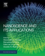
Liposomes are aqueous vesicles surrounded by concentric phospholipid bilayers and can carry lipophilic, hydrophilic, and amphiphilic pharmaceuticals. Since they are primarily constituted by lipids, liposomes are biodegradable, nontoxic, and nonimmunogenic compounds [62]. Fig. 4.5 illustrates the structure of a liposome and the appropriate locations for incorporation of pharmaceuticals with the different characteristics cited.

Figure 4.5 Structure of a single-layer liposome indicating the regions where pharmaceuticals with different characteristics can be incorporated.
Since they are constituted by a double lipid layer, liposomes are very similar to biological membranes. This characteristic promotes the permeation and transportation of pharmaceuticals through cell membranes and protects the drug from degradation by the reticuloendothelial system [63].
Liposomes are formed in water by the aggregation of amphiphilic molecules (usually phospholipids) that contain a polar group and an aliphatic chain. This process forms bilayers that close, forming an internal aqueous compartment. Volume 1 of this work, Chapter 2 (Supramolecular Systems), describes the primary methods of liposome preparation.
Liposomes are considered excellent delivery systems because of their structural flexibility, and they represent the most clinically established system for delivery of cytotoxic pharmaceuticals, genes, and vaccines [57]. There are countless medications on the market that use this delivery system [55,56,67], and many researchers have studied the possibility of using liposomes to carry pharmaceuticals for the treatment of Alzheimer’s disease [32,59]. However, improvements are still needed before liposomes can be used in the treatment of neurodegenerative diseases because their short half-life and low stability represent disadvantages yet to be overcome [58,59].
4.3.2.2.3. Biodegradable nanoparticles
Biodegradable particles and capsules at the nano- and micrometric scale are considered an alternative to the use of liposomes, as they allow a larger dose of pharmaceutical to be encapsulated and greater control of its delivery process. These advantages are provided by the large variety of materials available and the different methods for their preparation [68].
Microparticles are small, solid, and spherical particles with sizes that range from 1 to 1000 μm, while nanoparticles are drug carrier systems with diameters smaller than 100 nm. The term “nanoparticle” includes nanocapsules and nanospheres, which differ according to their composition and structural organization. The capsules are formed by a polymeric layer around an oily nucleus. The pharmaceutical can be dissolved in this nucleus and/or adsorbed in the polymeric wall. In turn, the spheres, which did not contain oil, are formed by a polymer matrix where the pharmaceutical of interest can be retained or absorbed [69]. Fig. 4.6 shows an illustration of these two systems, containing molecules of an incorporated medication.

Figure 4.6 Schematic illustration of nanospheres and nanocapsules with incorporated pharmaceuticals.
Once these nanosystems reach the target tissue, the drug starts to be released by processes, such as desorption, diffusion through the polymeric matrix, or erosion of the particle [56]. Nanoencapsulated drugs are more protected against degradation and phagocytosis by the reticuloendothelial system [58].
Countless studies have been performed to develop formulations of encapsulated pharmaceuticals, including for the treatment of CNS disease. For example, Rivastigmine, a drug used in the treatment of Alzheimer’s disease, was incorporated in poly(n-butyl cyanoacrylate) nanocapsules coated with polysorbate-80. According to the results of that study, when the encapsulated drug was employed, there was an increase in the availability of the pharmaceutical in the brain relative to the free form. This caused a decrease in the side effects of the drug, demonstrating the efficacy of the nanosystem [70].
In other studies, Tacrine, a medication also indicated for the treatment of Alzheimer’s disease, and a Bromocriptine, commonly used in the treatment of Parkinson’s disease, were nanoencapsulated in poly(n-butyl cyanoacrylate) coated with polysorbate-80 and poloxamer-188, respectively [58,71]. Similar to the previously cited study [70], a considerable increase was observed in the availability of the pharmaceuticals in the brain relative to the administration of their nonencapsulated free forms. Also according to these reports, Ritonavir (a medication used to treat HIV infections and AIDS) was carried by conjugated nanoparticles toward specific peptides for the treatment of neurological pathologies associated with HIV. These results indicated that nanosystems described previously are able to cross the HEB, providing an accumulation of the pharmaceutical in the CNS that is approximately 800 times higher than the conventional form of administration. Additionally, the use of this nanoencapsulated form increased the bioavailability of the pharmaceutical, maintaining the therapeutic concentration of the drug for up to 4 weeks [72].
Thus, nanoparticles represent an excellent alternative for the encapsulation of pharmaceuticals, especially for the diagnosis and treatment of neurological diseases, where crossing the HEB is the limiting factor in ensuring the appropriate concentration of the pharmaceutical in the CNS.
4.3.2.2.4. Micelles
Micelles are colloidal dispersions composed of particles with sizes between 5 and 100 nm [56]. These nanosystems have drawn much attention in drug delivery studies, largely due to their capacity to carry hydrophobic molecules and reach specific tissues. They are structures formed by an aggregate of molecules (generally surfactants or polymers) dispersed in a liquid [73]. Micelle preparation methods are discussed in Chapter 2 (Supramolecular Systems) in volume 1 of this work.
In regards to their geometry, micelles can be globular, ellipsoid, or layered, depending on their constitution and the conditions of the solution, such as concentration, temperature, pH, and ionic force. Micelles have specific characteristics, such as thermodynamic stability, and their “shell-core” structure imitates certain natural delivery systems, which promotes the absorption and distribution of the encapsulated medication. In comparison to liposomes, micelles are much smaller and can provide a more efficient method for guiding the pharmaceutical to the target site [56,58].
Due to their hydrophilic layer, micelles are not easily captured by macrophages from the reticuloendothelial defense system, instead protecting the incorporated drug from rapid degradation [59]. Micelles can solubilize lipophilic substances in their nucleus, hydrophilic substances adsorbed on their surface, and substances with neutral polarity (amphiphilic) along the molecules that form their structure [73]. Fig. 4.7 illustrates the structure of a micelle and the sites for the solubilization of substances with different polarities.
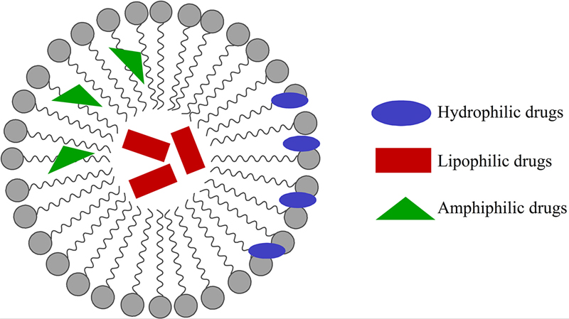
Figure 4.7 Structure of a micelle and substances with different polarities incorporated or adsorbed on its surface.
Although less extensive than some of the methodologies discussed previously, the use of micelles for the incorporation of drugs used in the treatment of CNS diseases has also been under study. For example, the effects of Haloperidol (a neuroleptic drug) in both its free form and when incorporated in Pluronic P85 micelles were compared, revealing greater effectiveness of the form incorporated into the micellar system [74].
Doxorubicin is an important drug used for the therapy of different types of tumors. However, it is not able to cross the HEB, so it cannot be used for the treatment of brain tumors. Nevertheless, a study has demonstrated that when this pharmaceutical is carried by polysorbate-80 micelles, considerable concentrations of the drug were detected in the brain [75].
Therefore, exploration of the use of micelles to release pharmaceuticals or diagnostic agents in the CNS could be a promising alternative for the development of efficient and versatile formulations.
4.3.2.2.5. Dendrimers
The word dendrimer derives from the Greek dendron, which means tree. Dendrimers are structures on the order of 5–20 nm, formed by radial growth in layers from a polyfunctionalized core, where the number of monomer units incorporated in each layer is successively doubled or tripled in relation to the previous cycle (Fig. 4.8). The resulting structure has a large number of ramifications and functional agglomerations on its surface [76].
Figure 4.8 Illustration of a dendrimer containing molecules of a pharmaceutical. Adapted from P. Kesharwani, K. Jain, N.K. Jain, Dendrimer as nanocarrier for drug delivery, Prog. Polym. Sci. 39 (2) (2014) 268–307, Copyright (2014) [77].With kind permission from Elsevier Publisher.
Encapsulation of a medication in dendrimers is performed through Van der Wals interactions and/or hydrogen bonds [56]. The advantage relative to other synthetic polymers is high molecular uniformity and high predictability of the molecular mass, size, and functional groups, providing more safety in relation to the composition and quantity of the incorporated pharmaceutical [78].
Some dendrimers are being used in biomedicine as cardiac markers for rapid diagnosis of heart attack, as a tool to promote in vitro gene transfection and as strategic biological sensors of anthrax or botulinum toxin [78]. There are also studies for the application of this nanosystem in the treatment of cancer [34,35].
In the specific case of neurological diseases, Kannan et al. [76] treated rabbits suffering from induced cerebral palsy using N-acetyl cysteine dendrimers. The drug was able to cross the HEB and reach the cells responsible for the inflammatory process involved in this disease, improving motor conditions and decreasing inflammation. Therefore, dendrimers also represent a nanosystem that could be used to carry drugs with neurological applications.
4.3.2.2.6. Nanosponges
Nanosponges represent a new class of controlled release system. These particles can carry hydrosoluble substances and promote the dispersion of certain lipophilic substances in the water. They are tiny structures with dimensions comparable to a virus and take the form of a mesh that is often produced from a type of biodegradable polyester (alginate, for instance), with segments that form spheres or cavities where the medication is incorporated [79] (Fig. 4.9).

Figure 4.9 Illustration of a nanosponge with incorporated medication.
Compared to other nanosystems, nanosponges are more soluble in water as well as in organic solvents. They have high porosity, are atoxic, and are stable at temperatures of up to 300°C. The only disadvantage of this system is that only small molecules can be incorporated [64].
Nanosponge-based formulations can be administered through oral, parenteral, topical, and inhaled routes. Studies performed with medications that are already commercialized in their free forms are being performed with the same compounds incorporated in nanosponges. These include Paclitaxel [80] and Tamoxifen [81] (both used for the treatment of cancer), Dexamethasone (antiinflammatory/antiallergy), and Itraconazole (antifungal) [82] as well as vaccines, antibodies, proteins, and enzymes [64]. To date, there are no reports of studies on the application of this system in neurological diseases, but they certainly have great potential to be explored.
Thus far, this chapter has briefly described the contributions of nanotechnology to the field of pharmacology that are promoting important breakthroughs in different areas of medicine, such as neurology. However, given the complexity of biological systems, the development of new pharmaceuticals and the study of their mechanisms of action still depend on the contribution of other fields of study, including informatics.
4.4. Computational Resources in Nanomedicine
From a broad perspective, informatics is itself the application of information and computational science methods to collect, analyze, and apply data in a specific situation. The use of prefixes in informatics, such as bioinformatics, ecoinformatics, and neuroinformatics, became the standard descriptor for the application of these methods to study problems within a specific field or subject. Likewise, computational science is the use of advanced computational techniques and sophisticated algorithms to position and solve problems. Here, the use of descriptive words in n-computational, such as in computational biophysics, refers to the use of computational methods within the field or subject of biophysics.
In the last two decades, the exploration of large amounts of data began to combine oriented experimental data with computer science and methods that use massive computational networks and state-of-the-art scientific tools as well as social network system technologies [83–85]. Pioneer projects to sequence and analyze the human genome [86] and the Xylella fastidiosa genome [87] are excellent examples of this combination of sciences and can demonstrate how computational sophistication and coordination of domain expertise can unite to overcome great scientific challenges (Fig. 4.10).

Figure 4.10 Xylella fastidiosa and its genome: a comprehensive view of the biochemical processes involved in its pathogenicity and survival in the xylem host.
The main functional categories are shown in bold, and the bacterial genes and gene products related to that function are organized in sections [87]. Reproduced from A.J.G. Simpson, F.C. Reinach, P. Arruda, F.A. Abreu, M. Acencio, et al., The genome sequence of the plant pathogen Xylella fastidiosa, 406 (6792) (2014) 151–157, Copyright (2014) [87]. With the permission of Nature Publishing Group.
The main functional categories are shown in bold, and the bacterial genes and gene products related to that function are organized in sections [87]. Reproduced from A.J.G. Simpson, F.C. Reinach, P. Arruda, F.A. Abreu, M. Acencio, et al., The genome sequence of the plant pathogen Xylella fastidiosa, 406 (6792) (2014) 151–157, Copyright (2014) [87]. With the permission of Nature Publishing Group.
4.4.1. Computational Neuroscience, Neuroinformatics, and Neurobiophysics
Computational neuroscience is an interdisciplinary science that unites different areas of neuroscience, computer science, applied physics, and mathematics. It is the primary theoretical method for investigating the function and mechanism of the nervous system. Computational neuroscience differs from the psychological connectionism and from the theories from disciplines such as machines learning, neural networks, and statistical learning theory, which emphasize the realist, functional, and biological description of neurons as well as their physiology and dynamics [83]. These models capture the essential characteristics of biological systems in many space–time scales, from membrane barriers and protein to chemical coupling in network oscillations, learning and memory. These computational resources are used to test hypotheses that could be directly verified through biological experiments. Currently, this field is rapidly expanding and leading to a revolution in neuroscience. Many pieces of software allow the realistic, rapid, and systematic in silico modeling of neurons, such as NEURON (http://www.neuron.yale.edu/). Furthermore, there are also methodologies of systems grouped by patterns of molecular expression and of neuronal population information, such as the “Brain Architecture Knowledge Management System” (BAMS—http://brancusi.usc.edu/bkms/), an interface created to integrate the results of different experiences and forms of neurological data [88].
Neuroinformatics combines neuroscience and informatics research, aiming to develop and apply advanced tools and methods essential for breakthroughs in the understanding of brain structure and function [89,90]. Using these methods, the principles of brain signal processing are extracted from biological systems and applied to the construction of artificial intelligent systems [83]. The construction of artificially intelligent systems generates valuable feedback on the sufficiency of neurobiological concepts. Therefore, neuroinformatics varies between the modeling of neurophysiological processes and the biologically inspired development of artificial information processing systems, such as many approaches to artificial neural networks [83,91].
Klopper (2005) [92] also defines neuroinformatics as an emerging area of informatics where microscopic implants are performed, allowing nanocomputers to improve the sensory input to the brain. This may enable the blind to see again, the deaf to hear again, and the paralyzed to recover their motion. Yamaji et al. [93] added that neuroinformatics is a new research tool that promotes the organization of data from neuroscience and databases using Internet technology, such as open access databases.
Generally, this subject comprises three main areas [84]:
1. data related to neuroscience and databases, increasingly capable of dealing with the complexity and organization of the nervous system, from the molecular to the behavioral level;
2. tools for acquisition, visualization, analysis, and distribution of data from the nervous system; and
3. simulation of theoretical and computational environments for the modeling and understanding of the brain.
The main objective of neuroinformatics is to help neuroscientists deal with the analysis, modeling, simulation, and management of information resources before, during, and after research (Fig. 4.11). As such these scientists need to have access to a common and remote environment able to provide tools for the organization of data and the storage of results. One example of this environment is the Visiome (http://platform.visiome.neuroinf.jp) Neuroinformatics platform, which approaches questions related to neuroscience and provides different portals for different fields of brain research [89,90,94].

Figure 4.11 Scientific applications of three-dimensional reconstruction: the tracking of axonal and dendritic morphology is often carried out for many purposes, such as the creation of neuronal identity, anatomical implementation, and realistic biophysical simulations of neuronal electrophysiology, as well as for performing morphometric and stereological analyses to determine the potential for connectivity. Data deposition in central databases makes reconstructions easily accessible for reuse in any of these applications as well as in data mining, education, and propagation [95]. Reproduced from R. Parekh, G.A. Ascoli, Neuronal morphology goes digital: a research hub for cellular and system neuroscience, Neuron 77 (6) (2014) 1017–1038, Copyright (2014) [95]. With permission from Elsevier Publisher.
Gillies and Willshaw [96] showed in their study that neuroinformatics techniques, particularly computational modeling, can provide a reliable method to unite and develop new pharmacological concepts and phenomena, such as the loss of dopamine, with electrophysiological characteristics. These precise models of brain dynamics can directly connect diseases, such as Parkinson’s disease, to effective treatments by simulating pharmaceutical input through the membrane channels of the brain as well as critical physiology and mechanisms of brain stimulation.
Complementing the previously cited concepts, neurobiophysics is the application of the basic principles of physics to the function of the nervous system. The methods of neurobiophysics are identical to those of other quantitative scientific fields and include the observation of phenomena under controlled conditions and the subsequent replication of those phenomena as well as the elaboration of models where the laws of physics can be applied and provide quantitative explanations of relevant observations [97].
A basic assumption in neurobiophysics is that all neuronal activity has a possible explanation based on the application of known laws of physics [98]. In this view, the morphological complexity of the neuron and the structural complexity of the neuronal interconnections are practical barriers for understanding the nervous system, and thus, neurobiophysics uses the computational sciences to investigate areas ranging from cell biology to complex neural network systems [97]. Table 4.1 shows the primary tools and resources for digital reconstructions that are currently available for the study of neural networks.
Table 4.1
Three-Dimensional Reconstruction Resources and Tools of Neuron Morphology [95]


Source: Reproduced from R. Parekh, G.A. Ascoli, Neuronal morphology goes digital: a research hub for cellular and system neuroscience, Neuron 77 (6) (2014) 1017–1038, Copyright (2014) [95]. With permission from Elsevier Publisher.
Platform: W, Windows; M, Mac; L, Linux; (C), C/C++; (J), Java; (I), plugin ImageJ; (O) others, that is, Matlab, Python, Lisp, or Fortran. Support: E, email; F, forum; G, guide (manual); L, discussion list; N, newsletter; T, tutorial; D, continuous development; A, actively maintained.
a Tools: Commercial.
b Tools: Open source, it is not open, all of the others, free.
c Tools: Morphological modeling (as opposed to electrophysiological).
4.4.2. Nano(bio)informatics
Nanotechnology is a means of integrating the biological and the artificial world [99] given that, in fact, nanoparticles and nanodevices can interact with biological systems. Therefore, from the perspective of computer science, nanotechnology can catalyze a paradigm change in the domain of Informatics. That is, the interaction of computational processes with biological, natural information from complex systems and materials designed at the nanoscale.
The application of nanotechnology to healthcare is known as nanomedicine [99]. Thus, the application of nanomedicine to the field of Informatics is called nanoinformatics. Nanoinformatics can be used with biomolecular approaches for diagnosis, preventive medicine, and monitoring and treatment of illnesses [99], in addition to promoting the creation of medical tools under the umbrella of nanobioinformatics. It is important to emphasize that while this discussion will only include its biomedical applications, nanoinformatics can also be related to other areas of nanotechnology.
Nanoinformatics has the purpose of building a bridge between nanomedicine and information technologies by applying computational methods to manage the information created within the domain of nanomedicine. Hence, nanoinformatics is simply the management of information, an approach for efficiently coordinating the resources available in scientific study and making predictions through molecular and biological modeling [100]. While bioinformatics is usually applied in the context of the analysis of DNA sequence data and the prediction of relationships between genetic mutations and diseases [101], Nanoinformatics is applied to organizing, interpreting, and making predictions of structures, physicochemical properties, environments, and applications associated with nanoparticles and nanomaterials, thereby promoting the evolution of medical fields in general [100,102,103]. These new applications are being born at a time when Genomic Medicine and customized treatment are still being developed and therefore hold great promise for Modern Medicine.
Currently, some fields are largely focused on the study of nanoinformatics [101]: nanoparticle characterization, nanotoxicology, modeling and simulation, imaging, terminology, ontologies and standardization, data integration and exchange, interoperability of systems, data and text mining for nanomedical research, connection of nanoinformation to computerized medical records, basic and translational research, creation of international networks of researchers, projects and labs, nanoinformatics education, and ethical matters.
Nanoinformatics education is still in its early phases. The subject of nanoinformatics was created with the support by the US National Science Foundation in 2007 and is therefore a very recent field of science. In Europe, this term was launched in 2008 in a project called ACTION-Grid and supported by the European Commission. Its purpose was to analyze the challenges surrounding and create a schedule for the development of this discipline, which is intrinsically connected to nanotechnology, biomedicine, and informatics [100,104].
In regards to nanotoxicology, nanoinformatics is present in studies related to the investigation of the toxicity of nanoscale products in natural systems (in vivo or in vitro). This type of information is fundamental for guaranteeing the safe use of nanomaterials for medical and pharmacological products [100] and is used in new clinical trials and nanoscale simulations generated by informatics. When speaking of toxicology at the nanoscale, it is important to emphasize that it refers to the nanometric size of particles as well as their unprecedented electrical, optical, chemical, and biological properties. A common methodology in nanoinformatics, with regard to toxicity, is the quantitative structure–activity relationship (QSAR), a process through which the chemical structure is quantitatively correlated to a well-defined process, such as the biological activity or chemical reactivity of a nanotechnology material. The development of QSAR generally requires information on well-characterized nanomaterial structures, experimental data that establish the activity of these materials within the environment where they are used, and a theoretical base founded on the verification of specific literature [105].
There are databases with information on toxicological effects, such as those from the National Institute for Occupational Safety and Health (NIOSH—https://www.cdc.gov/niosh/) and the oregon nanoscience and Microtechnologies Institute (ONAMI—http://www.onami.us/). In the near future, researchers will use nanoinformatics to model and simulate toxicity processes, relating them to the information of real patients from computerized medical reports to predict the human response to products, such as nanopharmaceuticals [102]. In Europe and the United States, similar initiatives, such as the Advancing Clinical Genomic Trials on Cancer (ACGT) and the Cancer Biomedical Informatics Grid (caBIG), have developed new approaches for data sharing, modeling, and simulation of drug delivery [102,106]. These initiatives are designed to accelerate the trials necessary to guarantee the efficacy and safety of pharmaceuticals. By using the advanced computational methods offered by nanoinformatics, doctors and researchers will be able to reduce the time taken by the laboratory–clinical practice to make nanopharmaceuticals available to patients [102].
4.4.3. Application of Informatics Methods and Tools to Nanomedical Data
In computer science, ontologies are used in artificial intelligence, web semantics, software engineering, and information architecture as a form to represent knowledge of the world in whole or part [107]. In turn, nanoinformatics ontologies can be considered models of data that represent a set of concepts within a domain, represented by nanomedical structures. These models contains countless concepts, nanomedical categories, and relationships that could be used as a controlled terminology for the management of data and analysis, allowing different investigation tasks, such as the registration of experimental data or the integration of heterogeneous data sources [108]. Considering the extensive medical, biological, and nanotechnological vocabulary, the ontologies provide a mechanism to standardize the representation of the nanomedical domain, which could be very useful to propagate information and knowledge, as shown in Fig. 4.12.

Figure 4.12 Medical nanoinformatics.
Currently, there are relevant initiatives for the development of nanomedical ontologies, such as nanoparticle ontology (NPO) [109,110], developed by researchers at the National Cancer Institute. In addition to the representation of domain knowledge, nanoinformatics could lead to another relevant challenge for nanomedicine: the management of large volumes of data and information generated by nanomedical research. This challenge demands methods to improve efficiency in the search and recovery of information, such as that provided by mining data in the medical literature or the location of information in large databases to access new discoveries and other information on reactions, chemical properties of nanoparticles, nanomaterials, nanotoxicity, and physical interactions of nanomaterials.
Based on the history of research and interdisciplinary cooperation, there is a clear potential for synergy among all areas to which computational nanoscience could contribute. In the not too distant future, this junction of disciplines could improve our current knowledge of medicine and, especially, could lead science to find currently unknown cures for many diseases.
Abbreviations
AFM Atomic force microscopy
CNS Central nervous system
DNA Deoxyribonucleic acid
HEB Hematoencephalic barrier
MS Multiple sclerosis
NMO Neuromyelitis optica
NPO Nanoparticle ontology
QSAR Quantitative structure-activity relationship
RNA Ribonucleic acid
SEM Scanning electron microscopy
Glossary
Biocompatible The presence of a material in an organism does not cause harmful reactions in the organism. For example, when the body adapts to an implant by forming a collagen capsule. In this case, the organism recognizes the implant as an “intruder”, but its presence does not cause harmful effects.
Biodegradable A material with biocompatible characteristics and that creates minimal organic interference because it is degraded by biological processes.
Excitotoxicity Toxicity due to excessive and nonnatural excitation of a nervous cell by a neurotransmitter. This is common in pathologies such as Parkinson’s disease and often causes cell death.
Pharmacodynamics The physiological effects of pharmaceuticals in an organism, the mechanism of action, and the relationship between the pharmaceutical concentration and effect.
Pharmacokinetics The “path” taken by a medication through an organism. It includes absorption, delivery, biotransformation, and excretion.
..................Content has been hidden....................
You can't read the all page of ebook, please click here login for view all page.